Foot function: an evidence review and proposed new framework

Our knowledge of foot function underpins our management of people with lower limb pain and injury and strategies for improving lower limb performance. Advances in technology, such as biplanar video-radiography, musculoskeletal imaging and intramuscular electromyography, has enabled investigation of foot function more rigorously than previously possible.
In 2023 Behling and colleagues published a review article1 that evaluated biomechanical theories of foot function enabled by new available data acquired with technology that early researchers did not possess. They conducted a literature review to describe the historical emergence of foot function theories, describe the key theories that attempt to explain human foot function and summarised the evidence for each theory. The Quality Assessment Tool for Studies with Diverse Designs was employed to determine the quality of studies and provide an overall score to represent the level of confidence in evidence (Score <14 = weak level of confidence; Score 14-26 = moderate level of confidence; Score >26 = strong level of confidence). Based on the findings they proposed a new framework for describing foot function that is consistent with current evidence.
Their review challenges us to reframe our understanding of human foot function and consider the implications this has for the clinical setting.
If you’ve ever wondered how much of what we have historically believed about foot function is supported by evidence, this blog is for you!
Discover our brand new Foot and Ankle Course
The following topics will be covered in this blog:
- Review of the evidence for foot function theories
- Mobile adaptor-rigid lever paradigm
- Arches of the foot
- Midfoot break
- Midtarsal locking
- (Hyper) Pronation
- Windlass mechanism
- Arch spring mechanism
- Key findings of the current evidence related to foot function
- Clinical implications
Review of the evidence for foot function theories:
Behling and colleagues’ review of the current scientific literature1 revealed that many of the theories that have historically underpinned our description of foot function were hypotheses based on assumption, not observation or experimental evidence. It is important to acknowledge that at the time many theories were proposed, existing methodologies often did not allow for empirical testing. However, considering technological advances that have made examinations feasible, existing evidence suggests that many of these previously held beliefs are not supported by scientific evidence.

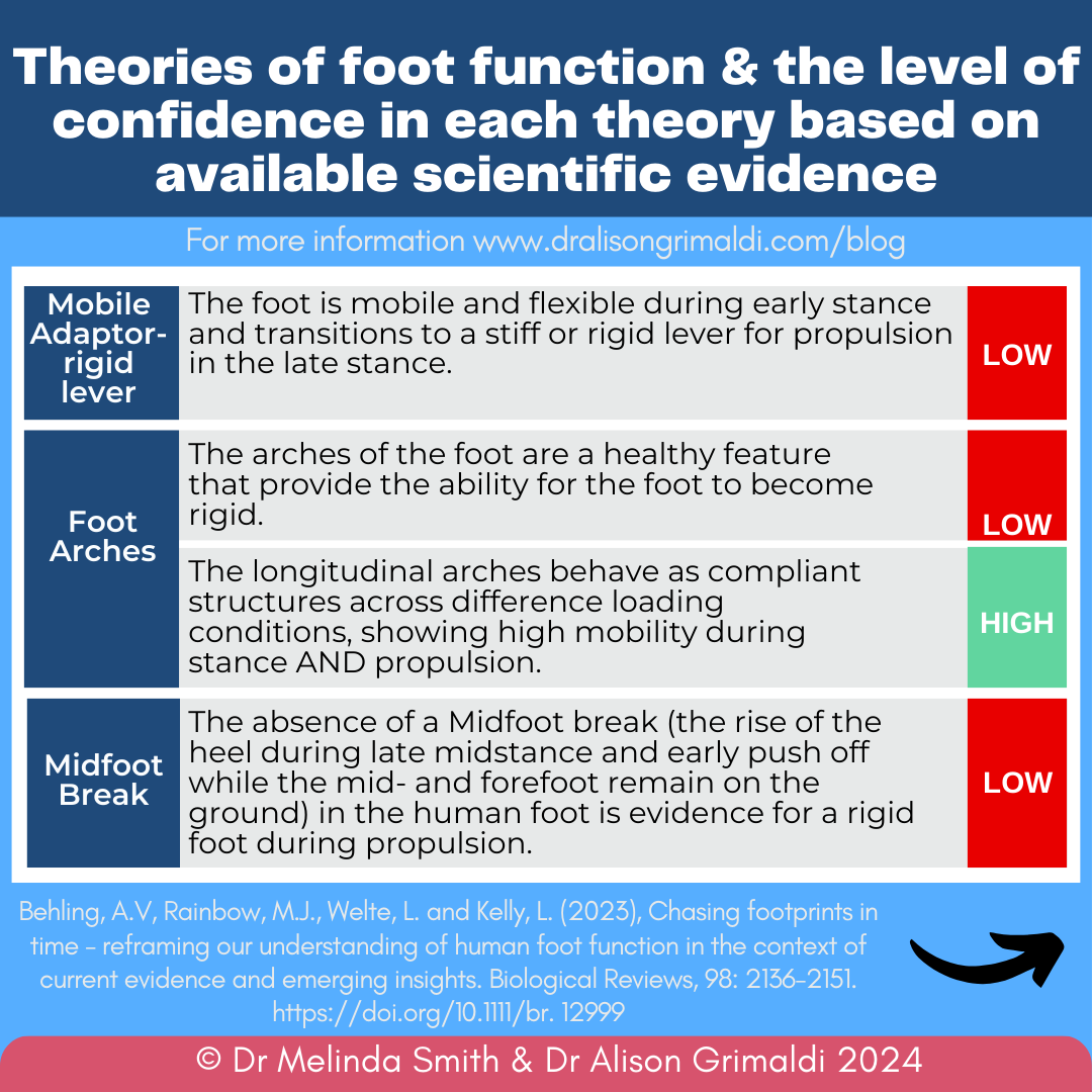

Let’s examine each theory in more detail.
Mobile adaptor-rigid lever paradigm
This theory describes a mobile and flexible foot during early stance that transitions to a stiff or rigid lever for propulsion in late stance. In the early 1900s the foot was described as immobile and rigid with authors of the time arguing that this was required to provide a stable support for the body during gait.2 In early gait analysis studies,3,4 rearfoot and midfoot pronation were reported during early stance and were thought to relate to attenuation of initial impact. Consequently, a mobile foot was considered a critical component of shock attenuation. A review of these early studies revealed that the mobile adaptor-rigid lever paradigm was a hypothesis based on assumptions, not observation, and there was no experimental evidence to support that the foot transitioned from a mobile adaptor to a rigid lever during gait. A large contributor to the lack of experimental evidence was the limitation in methodologies, which were not possible until recent years.
Discover our Foot and Ankle Course

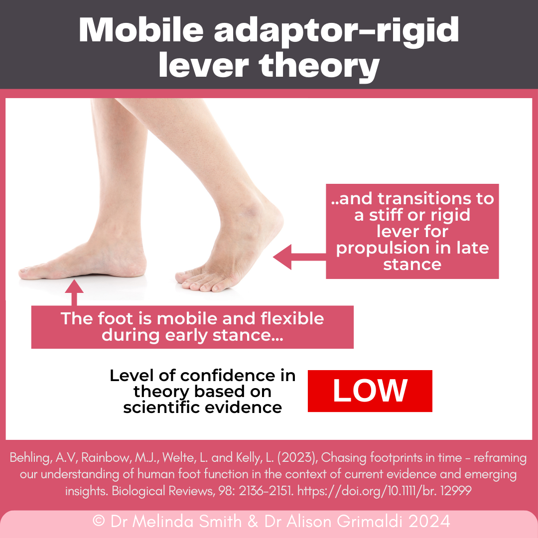
The mobile adaptor-rigid lever paradigm led to the creation of several theories. We will now discuss each of these and the related evidence.
Arches of the foot
The human foot has three arches: the medial longitudinal arch, lateral longitudinal arch and transverse arch. The arches of the foot have historically been considered a ‘healthy’ feature that enabled a rigid foot. The medial longitudinal arch in particular was considered a distinguishing feature of the human foot when compared to the flat and flexible primate foot. This observation contributed to the proposition that the (longitudinal) arches provided the human foot an ability to become rigid.
Since these early descriptive/observational studies that considered the arches as critical to bipedal function,2,5 several experimental studies have examined longitudinal arch behaviour. These more recent experimental and quantitative studies, with higher confidence in their scientific quality, have reported substantial mobility in the human foot during early stance and propulsion.6-11 So, although the longitudinal arches are a unique feature of the human foot, this does not prevent them from deforming underload and arch height has not been correlated with arch stiffness.

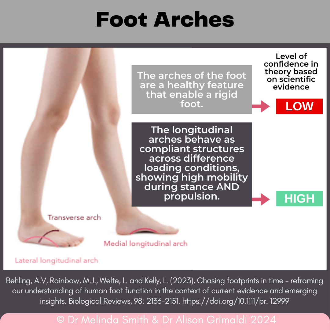
There has been less examination of transverse arch behaviour. Although there is a lack of in vivo validation, in vitro studies and biomechanical modelling suggest that the transverse arch ligaments stiffen the longitudinal arch by 40%.12 This suggests there may be an interplay between transverse and longitudinal arch stiffness, but further examination is required.
Midfoot break
Midfoot break describes the rise of the heel during late midstance and early push-off while the midfoot and forefoot remain on the ground. It was thought to be a characteristic feature of the non-human primate foot. Early observation of the absence of a midfoot break in the human foot was considered evidence for a rigid foot for propulsion.2,13
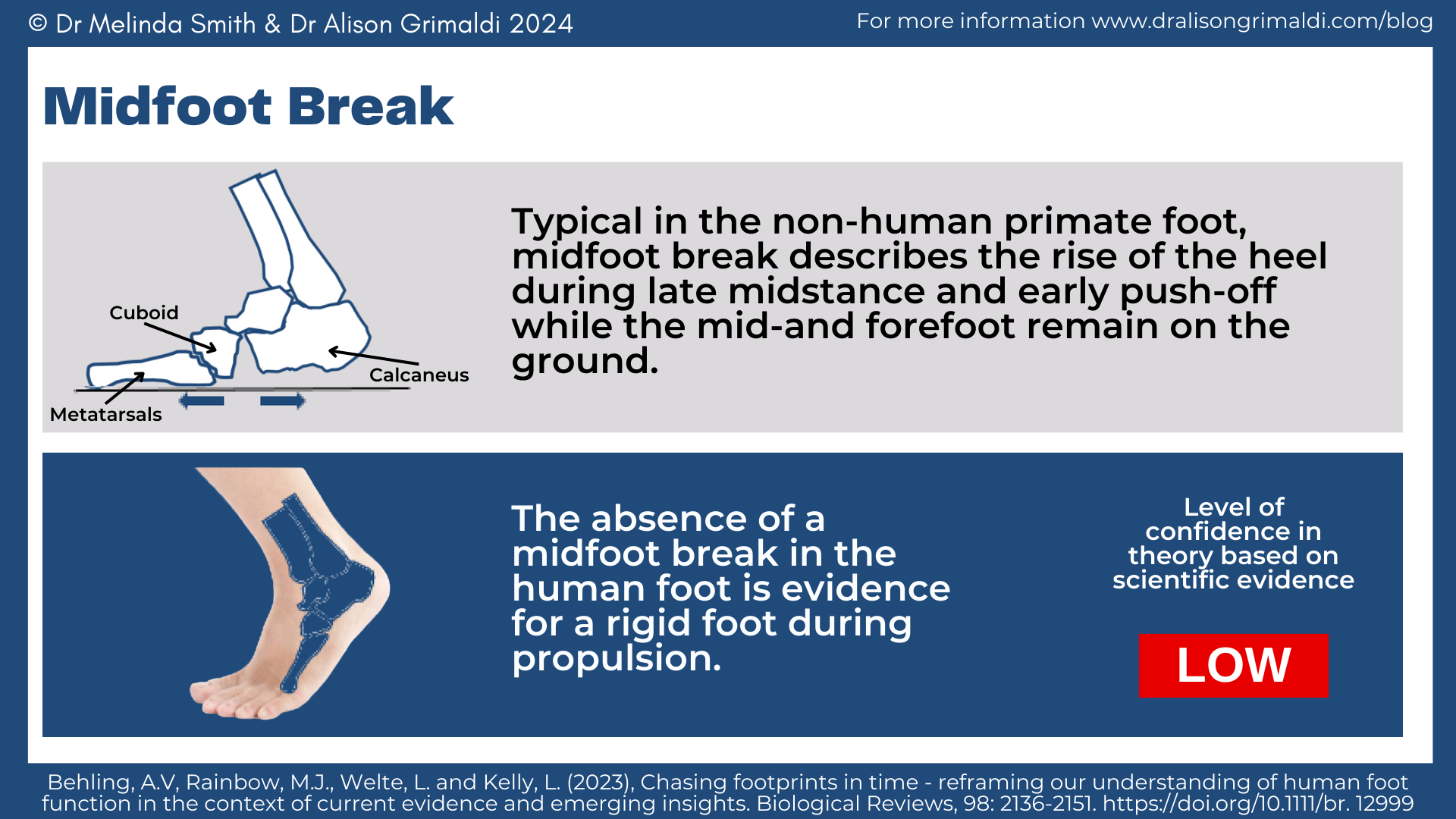

Since these early observations, quantitative measurements from several biomechanical studies in the 2000s onwards have demonstrated that the midfoot break occurs as a continuous trait in healthy human feet.14,15 Furthermore, they reported that human feet with a midfoot break were still able to successfully push-off during walking.15
Midtarsal locking
Midtarsal locking describes the talo-navicular and calcaneo-cuboid joints (i.e. the midtarsal joints) become immobilised (‘locked’) during the transition to push-off because of calcaneal inversion. The ‘locking’ was explained by two mechanisms – (1) a screw-like interaction of the cuboid rotating into the distal calcaneal facet as the calcaneus inverts, and (2) with calcaneal inversion the axes of the talo-navicular and calcaneo-cuboid joints are misaligned/converge.16-18 Early studies describing these mechanisms were descriptive in nature and a lack of suitable measurement tools prevented evaluation of the midtarsal locking hypothesis.
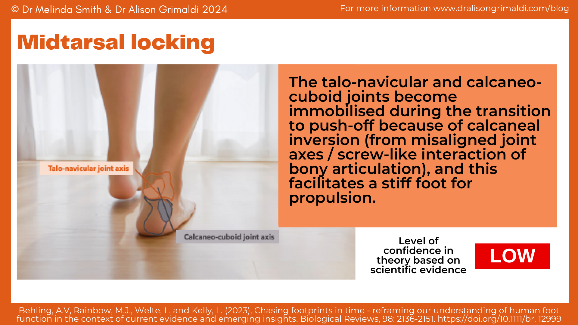

Following advances in technology and biomechanical methods, more recent bone pin studies have demonstrated that midtarsal locking does not exist.15,19-22 Dynamic cadaver simulations of gait demonstrated that the relative orientation of the midtarsal joint axes remained constant during foot flat and were slightly more aligned (not misaligned/converged) at push-off. Furthermore, similar amounts of midtarsal movement during landing (supposedly when the midtarsal joint is unlocked) and push-off (supposedly when the midtarsal joint is locked) have been reported. Based on current available evidence, Behling and colleagues concluded that midtarsal locking as a hypothesis should be rejected, as well as the general notion that the midfoot is immobile during push-off.
(Hyper) Pronation
Pronation is a triplanar movement that consists of rearfoot eversion, forefoot abduction and foot dorsiflexion relative to the talus. In the early 1900s pronation was associated with an unlocked midtarsal joint that, whilst desirable for shock absorption at heel contact, was thought to disrupt the transition to a rigid lever for propulsion.
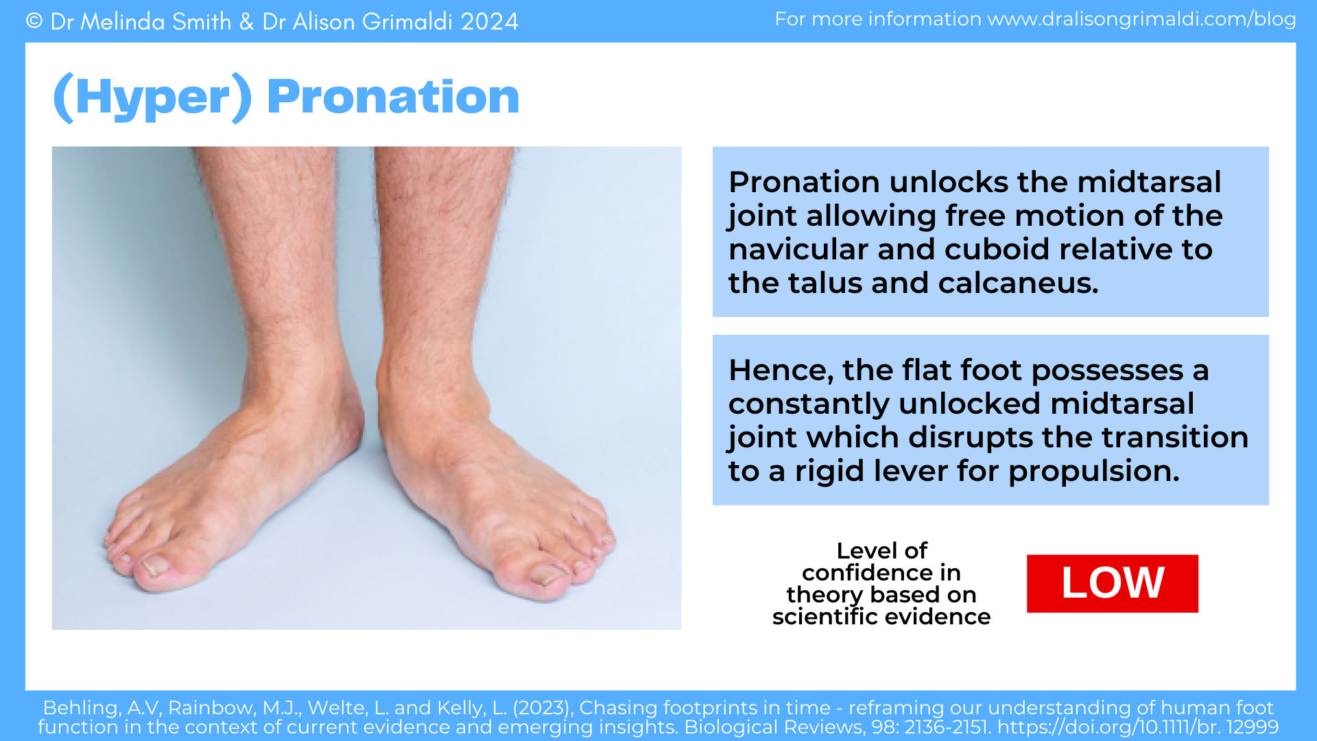
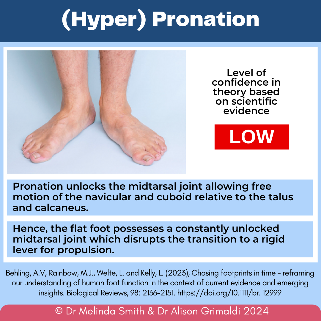
Historically, a pronated flat foot was considered an undesirable feature linked to pain and injury. The early reports that informed this belief were descriptive and observational with low levels of confidence in their conclusions. Investigations since then generally suggest that flat feet are a healthy variation of foot morphology among humans.
Studies have demonstrated that pronation is a naturally occurring movement23,24 that has not been consistently linked with injury.25-27 Biomechanical studies using 3-dimensional motion capture, dynamic ultrasound imaging and intramuscular electromyography during walking reported that the spring-like function of the foot occurred regardless of foot posture.28,29 These studies showed that subtalar joint pronation during early stance allowed energy to be stored in the tendon of the tibialis posterior muscle and subsequently returned during propulsion as the subtalar joint supinates and the tibialis posterior tendon recoils. Foot mobility may therefore be an important contributor the human foot function rather than a problem linked to pathology. This also highlights the important role of the extrinsic muscles in supporting foot function.
Windlass mechanism
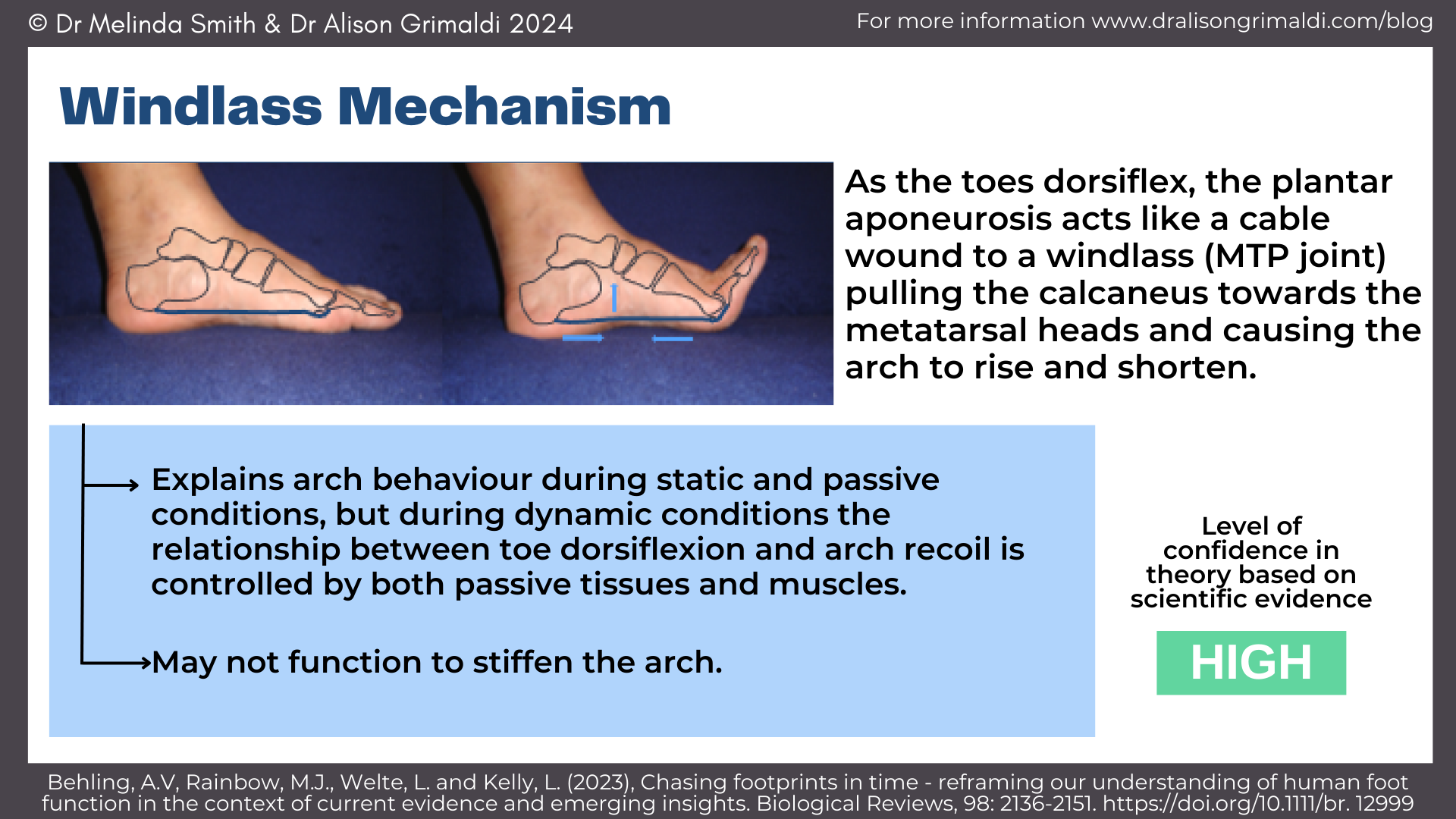
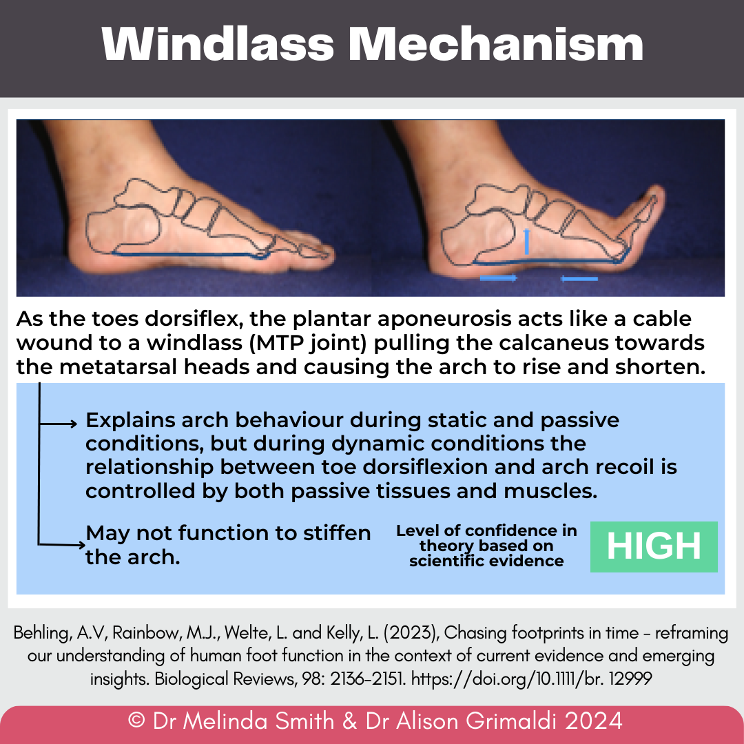
First proposed by Hicks in 1954,30 the windlass mechanism describes that as the toes dorsiflex the plantar aponeurosis acts like a cable wound to a windlass (the metatarsophalangeal joint) pulling the calcaneus towards the metatarsal heads and causing the arch to rise and shorten. This became one of the explanations of how the foot stiffens and transitions from a mobile adaptor in early stance to a rigid lever at push-off. The windlass mechanism became synonymous with stiffness and structural integrity. Presumably this was based on the early assumptions that a higher arch was indicative of a stiffer foot, and that the plantar aponeurosis was a non-straining cable (hence tension developed in the plantar aponeurosis increasing arch stiffness).
Early cadaver investigations at the time, as well as more recent in vivo studies, support the presence of a windlass mechanism.10,11,30,31 Whether this is associated with a stiffer foot is less clear with some studies reporting a more compliant (less stiff) foot when the windlass was engaged.10,32 Although there is minimal elongation in the plantar aponeurosis as the heel rises (supporting a windlass mechanism), it shortens rapidly as the arch recoils in mid to late stance suggesting a decline in tension.33,34
In recent years, further insight has come from additional biomechanical studies that have highlighted the role of muscles in stiffening the foot.8,9,35,36 Evidence suggests that the windlass mechanism explains arch behaviour during static and passive conditions, but during dynamic conditions the relationship between toe dorsiflexion and arch recoil is controlled by both passive tissues and muscles.
Arch spring mechanism
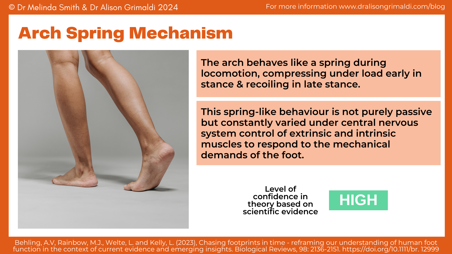
In 1987 Ker and colleagues proposed that the arch behaved like a spring during locomotion.37 They described that the arch compresses under load as ground reaction force increases in early stance and then recoils as the ground reaction force subsides in late stance. This was proposed to contribute to the efficiency of the foot with elastic strain of the plantar aponeurosis and plantar ligaments allowing energy to be stored and recycled within the foot with recoil. Early scientific studies in vitro and in vivo supported the hypothesis. 37
However, what more recent studies have added is insight that the foot does not display pure spring-like behaviour – some of the energy absorbed within the foot is not returned with arch recoil. Kelly and colleagues7 suggested that the intrinsic muscles of the foot may actively control the foot-spring function. Using intramuscular electromyography and 3-dimensional motion analysis, they reported that during walking and running the intrinsic foot muscles actively lengthen in early-midstance and shorten in late stance. In a subsequent study8, the same group of authors used a peripheral nerve block to remove active contribution from the intrinsic muscles of the foot. They reported that without intrinsic foot muscle activation, power output from the foot declined. Current evidence therefore supports an arch spring mechanism exists, but that it is not purely passive.
In recent years, biomechanical studies have expanded on these findings to demonstrate that the foot behaves as a damper when energy needs to be dissipated to decelerate the body and can produce power when the body needs to accelerate.35,38 Forces produced by the extrinsic and intrinsic foot muscles, acting in parallel to passive structures (plantar aponeurosis, plantar ligaments), enable mobility of the longitudinal arch which is vital to the versatile function of the foot.

Key findings of the current evidence related to foot function:
- The overarching mobile adapter-rigid lever paradigm has never been proven directly and evidence suggest that the foot never transitions from mobile in early stance to rigid in propulsion.
- The foot displays substantial mobility throughout the gait cycle (in early stance AND at push-off).
- Medial longitudinal arch height should not be associated with foot health.
- The notion that the shape of the foot (i.e. presence of a medial longitudinal arch) is important to the foot’s stiffening mechanism is not supported by evidence.
- Arches of the human foot exist on a continuum of mechanical structure (i.e. range of compliance-stiffness) which may be governed by the combination of bony configurations, ligaments, aponeuroses, muscles and tendon.
- Foot function is highly versatile. Evidence suggests that longitudinal arch mobility, rather than stiffness, is a key component enabling the versatile function of the human foot.
- The central nervous system governs the mechanical behaviour of the foot via control of extrinsic and intrinsic muscles that act in parallel and in series to passive elastic tissues (e.g. plantar aponeurosis and ligaments).
- The extrinsic and intrinsic muscles of the foot (under central nervous system control) can add or remove (dampen) energy to meet the requirements of the body and are therefore crucial to support the adaptive mechanical behaviour of the foot.
Clinical implications:
The review paper highlighted several gaps in the scientific evidence that have led to misconceptions in our understanding of foot function. In light of the current available evidence, we are urged to reconsider some theories that might underpin our clinical practice and reframe our conceptual understanding.

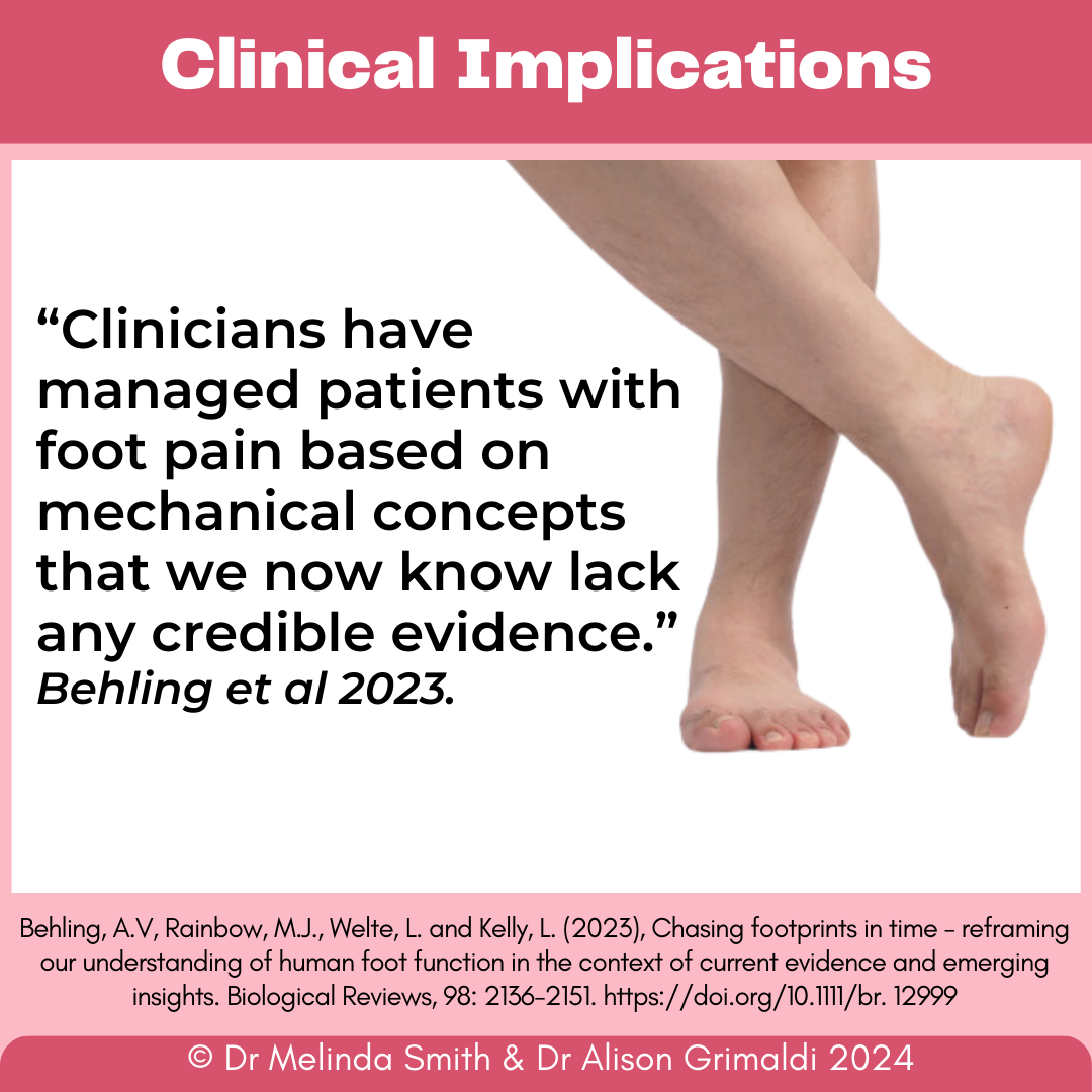
A review of the current evidence demonstrated the mobile adaptor-rigid lever paradigm was a hypothesis that was based on an assumption which has not been substantiated with subsequent research. Evidence suggests that the foot does not transition from mobile in early stance to rigid at propulsion. On the contrary, our feet display substantial mobility throughout the entire stance phase of walking i.e. during early stance AND at push-off. We need to stop describing the foot as a mobile adaptor that becomes a rigid lever. Rather, the foot supports body weight and facilitates propulsion as a dynamic system that is mobile throughout the gait cycle with longitudinal arch mobility (rather than stiffness) being a key component enabling the versatile function of the human foot.
The notion that the presence of a medial longitudinal arch is important to the foot’s stiffening mechanism is not supported by evidence. We should reconsider terminology that implies arch structure is related to a healthy foot. Although there may be some circumstances of concern, such as adult-acquired flat foot deformity, generally the evidence demonstrates that flat feet are a healthy variation of foot morphology among humans. We should avoid broadly associating longitudinal arch height with foot health or describing flat foot postures as inherently ‘bad’. Together with bone and joint morphology, the neuromechanical control and soft-tissue interactions contribute to foot function.
“The central nervous system delivers adaptive control, with the muscles of the foot adding or removing energy from the system as required to meet whole-body locomotory demands.” Behling et al 2023
If we think of the clinical implications of this quote from Behling and collegues1, the extrinsic and intrinsic muscles that control the foot underpin the highly versatile mechanical behaviour of the foot. Assessment and treatment of these muscles is therefore an important component in our management of people with lower limb pain and injury and to optimise lower limb function.
If you’d like to learn more about assessment and therapeutic exercise for the extrinsic and intrinsic foot muscles, consider our online course or face-to-face workshop “Mastering Movement of the Foot and Ankle”. You might also find the previous blog “Intrinsic muscles of the foot: small but mighty!” an interesting read.
We hope you’ve found this blog on foot function helpful!
We hope you’ve found this blog on foot function helpful!
Discover our Foot and Ankle Course
Check Out Some More Foot and Ankle Blogs:
References
- Behling AV, Rainbow MJ, Welte L, Kelly L. Chasing footprints in time - reframing our understanding of human foot function in the context of current evidence and emerging insights. Biol Rev Camb Philos Soc. Dec 2023;98(6):2136-2151. doi:10.1111/brv.12999
- Keith A. Hunterian Lectures ON MAN'S POSTURE: ITS EVOLUTION AND DISORDERS: Given at the Royal College of Surgeons of England. Br Med J. Apr 21 1923;1(3251):669-72. doi:10.1136/bmj.1.3251.669
- Inman VT. Human locomotion. Can Med Assoc J. May 14 1966;94(20):1047-54.
- Inman VT. The influence of the foot-ankle complex on the proximal skeletal structures. Artif Limbs. Spring 1969;13(1):59-65.
- Morton DJ. Evolution of the human foot. Am J Phys Anthropol. 1922;5(4):305-336. doi:10.1002/ajpa.1330050409
- Kelly LA, Cresswell AG, Racinais S, Whiteley R, Lichtwark G. Intrinsic foot muscles have the capacity to control deformation of the longitudinal arch. J R Soc Interface. Apr 6 2014;11(93):20131188. doi:10.1098/rsif.2013.1188
- Kelly LA, Lichtwark G, Cresswell AG. Active regulation of longitudinal arch compression and recoil during walking and running. J R Soc Interface. Jan 6 2015;12(102):20141076. doi:10.1098/rsif.2014.1076
- Farris DJ, Kelly LA, Cresswell AG, Lichtwark GA. The functional importance of human foot muscles for bipedal locomotion. Proc Natl Acad Sci U S A. Jan 29 2019;116(5):1645-1650. doi:10.1073/pnas.1812820116
- Farris DJ, Birch J, Kelly L. Foot stiffening during the push-off phase of human walking is linked to active muscle contraction, and not the windlass mechanism. J R Soc Interface. Jul 2020;17(168):20200208. doi:10.1098/rsif.2020.0208
- Welte L, Kelly LA, Lichtwark GA, Rainbow MJ. Influence of the windlass mechanism on arch-spring mechanics during dynamic foot arch deformation. J R Soc Interface. Aug 2018;15(145)doi:10.1098/rsif.2018.0270
- Welte L, Kelly LA, Kessler SE, et al. The extensibility of the plantar fascia influences the windlass mechanism during human running. Proc Biol Sci. Jan 27 2021;288(1943):20202095. doi:10.1098/rspb.2020.2095
- Venkadesan M, Yawar A, Eng CM, et al. Stiffness of the human foot and evolution of the transverse arch. Nature. Mar 2020;579(7797):97-100. doi:10.1038/s41586-020-2053-y
- Elftman H, Manter J. Chimpanzee and human feet in bipedal walking. Am J Phys Anthropol. 1935;20(1):69-79. doi:10.1002/ajpa.1330200109
- Bates KT, Collins D, Savage R, et al. The evolution of compliance in the human lateral mid-foot. Proc Biol Sci. Oct 22 2013;280(1769):20131818. doi:10.1098/rspb.2013.1818
- DeSilva JM, Bonne-Annee R, Swanson Z, et al. Midtarsal break variation in modern humans: Functional causes, skeletal correlates, and paleontological implications. Am J Phys Anthropol. Apr 2015;156(4):543-52. doi:10.1002/ajpa.22699
- Elftman H, Manter J. The Evolution of the Human Foot, with Especial Reference to the Joints. J Anat. 1935;70(Pt 1):56-67.
- Elftman H. The transverse tarsal joint and its control. Clin Orthop. 1960;16:41-46.
- Manter JT. Movements of the subtalar and transverse tarsal joints. The Anatomical record. 1941;80(4):397-410. doi:10.1002/ar.1090800402
- Bruening DA, Pohl MB, Takahashi KZ, Barrios JA. Midtarsal locking, the windlass mechanism, and running strike pattern: A kinematic and kinetic assessment. J Biomech. May 17 2018;73:185-191. doi:10.1016/j.jbiomech.2018.04.010
- Nester C, Jones RK, Liu A, et al. Foot kinematics during walking measured using bone and surface mounted markers. J Biomech. 2007;40(15):3412-23. doi:10.1016/j.jbiomech.2007.05.019
- Lundgren P, Nester C, Liu A, et al. Invasive in vivo measurement of rear-, mid- and forefoot motion during walking. Gait Posture. Jul 2008;28(1):93-100. doi:10.1016/j.gaitpost.2007.10.009
- Okita N, Meyers SA, Challis JH, Sharkey NA. Midtarsal joint locking: new perspectives on an old paradigm. J Orthop Res. Jan 2014;32(1):110-5. doi:10.1002/jor.22477
- McPoil T, Cornwall MW. Relationship between neutral subtalar joint position and pattern of rearfoot motion during walking. Foot Ankle Int. Mar 1994;15(3):141-5. doi:10.1177/107110079401500309
- Novacheck TF. The biomechanics of running. Gait Posture. Jan 1 1998;7(1):77-95. doi:10.1016/s0966-6362(97)00038-6
- Nielsen RO, Buist I, Parner ET, et al. Foot pronation is not associated with increased injury risk in novice runners wearing a neutral shoe: a 1-year prospective cohort study. Br J Sports Med. Mar 2014;48(6):440-7. doi:10.1136/bjsports-2013-092202
- Neal BS, Griffiths IB, Dowling GJ, et al. Foot posture as a risk factor for lower limb overuse injury: a systematic review and meta‐analysis. J Foot Ankle Res. 2014;7(1):55-n/a. doi:10.1186/s13047-014-0055-4
- Dowling GJ, Murley GS, Munteanu SE, et al. Dynamic foot function as a risk factor for lower limb overuse injury: a systematic review. J Foot Ankle Res. 2014;7(1):53-n/a. doi:10.1186/s13047-014-0053-6
- Maharaj JN, Cresswell AG, Lichtwark GA. The mechanical function of the tibialis posterior muscle and its tendon during locomotion. J Biomech. Oct 3 2016;49(14):3238-3243. doi:10.1016/j.jbiomech.2016.08.006
- Maharaj JN, Cresswell AG, Lichtwark GA. Subtalar Joint Pronation and Energy Absorption Requirements During Walking are Related to Tibialis Posterior Tendinous Tissue Strain. Sci Rep. Dec 20 2017;7(1):17958. doi:10.1038/s41598-017-17771-7
- Hicks JH. The mechanics of the foot. II. The plantar aponeurosis and the arch. J Anat. Jan 1954;88(1):25-30.
- Hicks JH. The mechanics of the foot. I. The joints. J Anat. Oct 1953;87(4):345-57.
- Sichting F, Ebrecht F. The rise of the longitudinal arch when sitting, standing, and walking: Contributions of the windlass mechanism. PLoS One. 2021;16(4):e0249965. doi:10.1371/journal.pone.0249965
- Caravaggi P, Pataky T, Gunther M, Savage R, Crompton R. Dynamics of longitudinal arch support in relation to walking speed: contribution of the plantar aponeurosis. J Anat. Sep 2010;217(3):254-61. doi:10.1111/j.1469-7580.2010.01261.x
- Williams LR, Ridge ST, Johnson AW, Arch ES, Bruening DA. The influence of the windlass mechanism on kinematic and kinetic foot joint coupling. J Foot Ankle Res. Feb 16 2022;15(1):16. doi:10.1186/s13047-022-00520-z
- Smith RE, Lichtwark GA, Kelly LA. The energetic function of the human foot and its muscles during accelerations and decelerations. J Exp Biol. Jul 1 2021;224(13)doi:10.1242/jeb.242263
- Smith RE, Lichtwark GA, Kelly LA. Flexor digitorum brevis utilizes elastic strain energy to contribute to both work generation and energy absorption at the foot. J Exp Biol. Apr 15 2022;225(8)doi:10.1242/jeb.243792
- Ker RF, Bennett MB, Bibby SR, Kester RC, Alexander RM. The spring in the arch of the human foot. Nature. Jan 8-14 1987;325(7000):147-9. doi:10.1038/325147a0
- Riddick R, Farris DJ, Kelly LA. The foot is more than a spring: human foot muscles perform work to adapt to the energetic requirements of locomotion. J R Soc Interface. Jan 31 2019;16(150):20180680. doi:10.1098/rsif.2018.0680

