Mastering the Calf Raise Test – More than just a Simple Lift

The calf muscles are a powerhouse of human movement. They are the primary plantar flexors of the ankle joint and making important contributions to lower limb function including walking, running and jumping. Their important contribution to human movement makes calf muscle function a common assessment in the clinical setting. The calf raise test (also known as the heel-rise test, single-leg heel raise, standing heel raise) has emerged as a widely used clinical tool for evaluating calf muscle-tendon unit performance. Counting the number of repetitions an individual can lift their heel off the ground might sound like a simple test, but there are many aspects of performing the test that can influence the result, and, additional insights beyond the count that are achievable.
The following topics will be covered in this blog:
- Calf muscle structure and function
- Assessing calf function using the calf raise test
- What is the calf raise test?
- Outcome measures from the calf raise test
- Factors that influence outcome of the calf raise test
- Interpretation of the calf raise test
- When to use the calf raise test
Register for the Upcoming Melbourne Workshop!
Want to learn more about assessment and training of foot & ankle function? Join us at our face-to-face workshop in Melbourne on February 8th. Register now so you can jump into the fantastic online learning content. Learn about the different aspects of foot and ankle function and the implications this has for muscle and movement assessment and therapeutic exercise prescription around the foot and ankle. Then dive into the practical elements during the workshop.
EARLY BIRD OFFER (Ends 7th December)
Calf muscle structure and function
Calf muscle structure
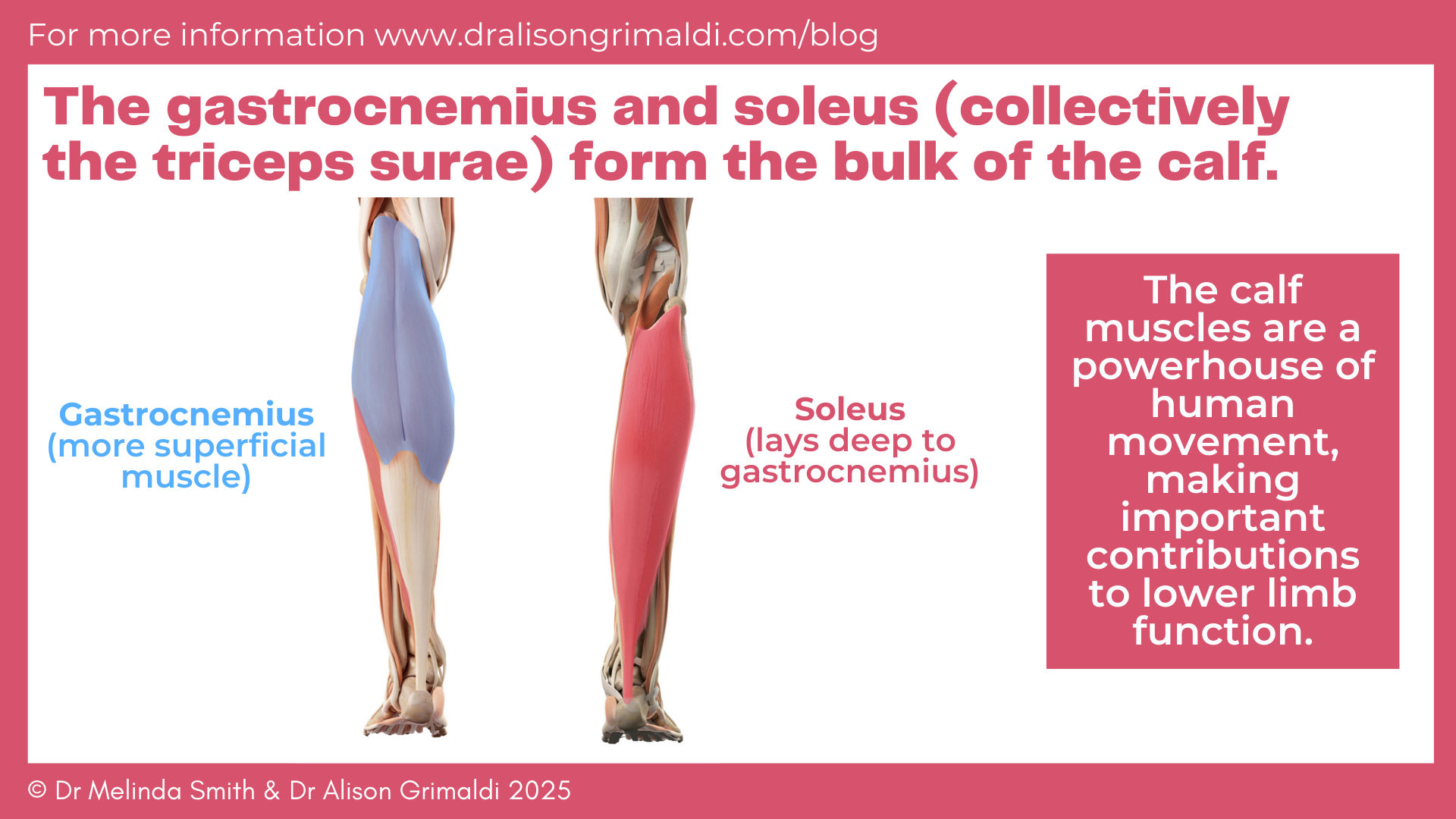
The gastrocnemius and soleus muscles (collectively known as the triceps surae) form the bulk of the calf (Figure 1 - below).1 The gastrocnemius muscle is more superficial and originates from two heads – one lateral and one medial – connected to the condyles of the femur.
The medial head originates on the posterior aspect of the distal femur just behind the adductor tubercle and above the articular surface of the medial condyle. The lateral head originates from the upper lateral surface of the lateral femoral condyle where it joins the lateral supracondylar line.
Both heads also arise from the underlying knee joint capsule. As the gastrocnemius muscle descends, about mid-calf, the fibres insert into a broad aponeurosis which gradually narrows and converges with the deeper soleus muscle to form the calcaneal (Achilles) tendon.
The soleus muscle is a large flat muscle under (deep to) the gastrocnemius muscle. It has a complex, three-dimensional structure consisting of a unipennate posterior part wrapped around a radially bipennate anterior part.2 It originates from the head and proximal end of the fibula, soleal line and middle third of the tibia, and a fibrous band which spans between the fibula and tibia (tendinous arch of the soleus).
The origin of the soleus muscle is aponeurotic with fibres that arise from the posterior and anterior surface of the aponeurosis. Inferiorly muscle fibres insert into a long distal central tendon that merges with the overlying distal tendon of gastrocnemius to form the calcaneal (Achilles) tendon. Proximally the soleus muscle is covered by the gastrocnemius, but distally it is broader than the tendon of the gastrocnemius and accessible either side of the tendon (Figure 1 - below).
Via the calcaneal (Achilles) tendon, both muscles insert on the calcaneus of the foot.
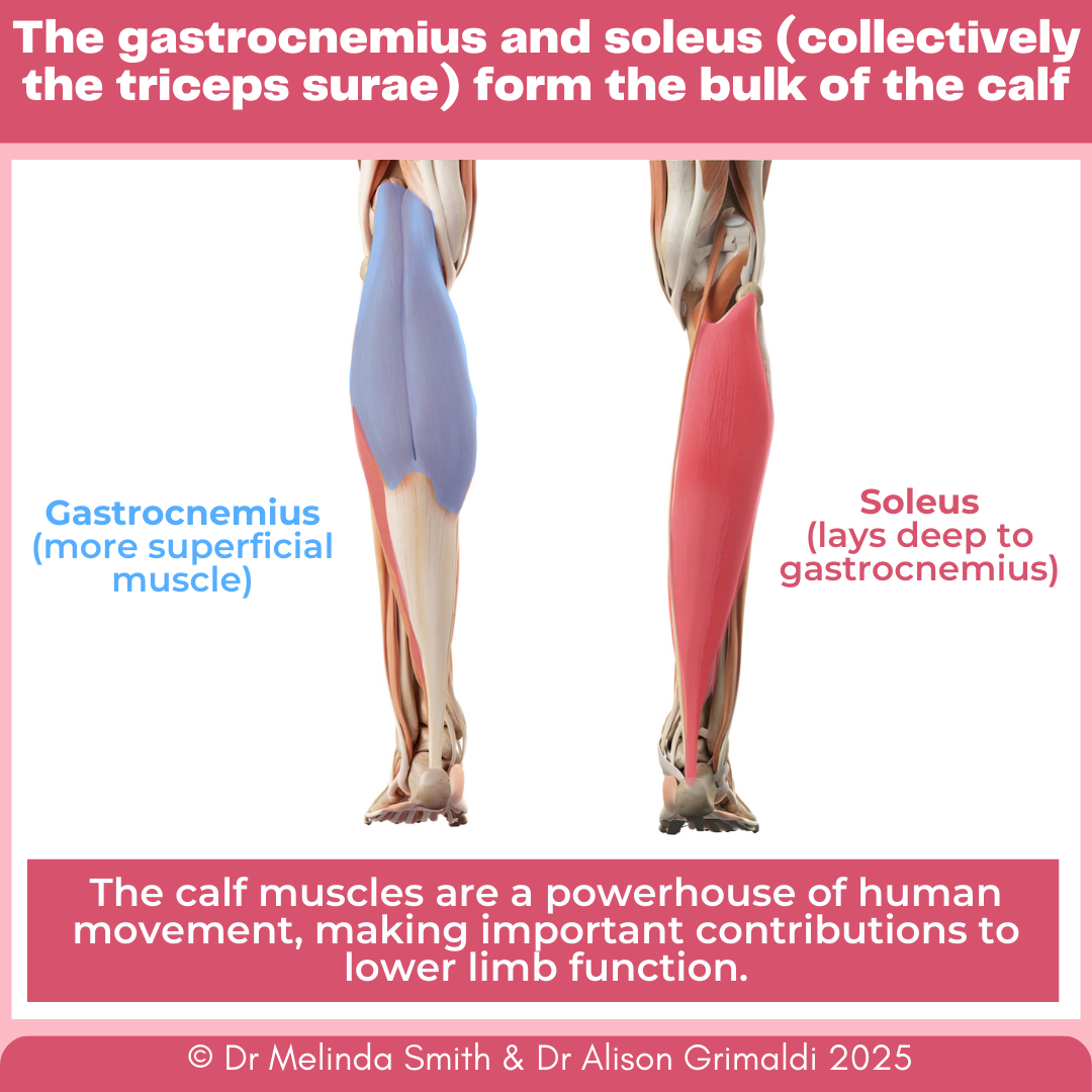
When compared with other major human lower-limb muscles, the gastrocnemius and soleus have unique structural designs in that both have short muscle fibers connected to the heel via a long and compliant Achilles tendon. This architectural arrangement has important functional implications – it allows the triceps surae complex to optimize biomechanical efficiency. The lengthy tendon absorbs and distributes much of the length changes during movement through its elastic stretch and recoil properties, while the muscle fibres maintain slower shortening velocities conducive to effective force generation.3
Calf muscle function
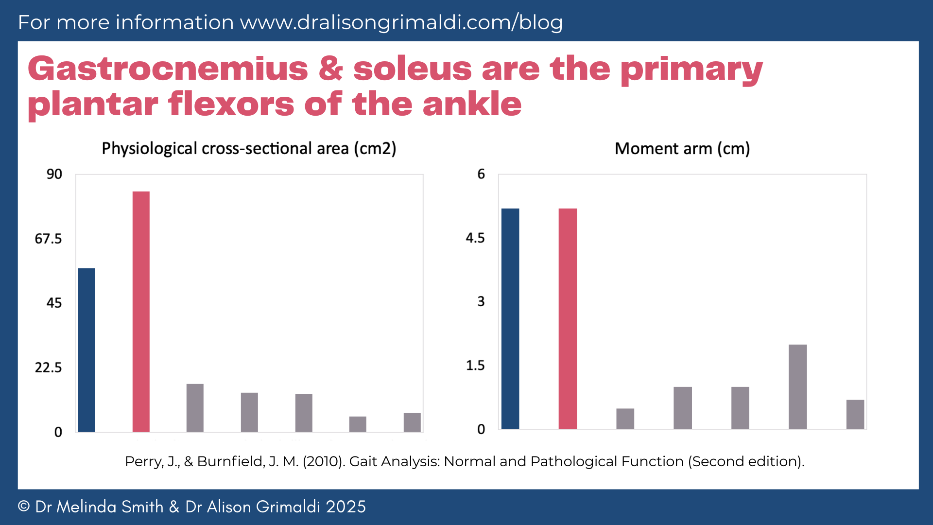
Seven muscles pass posterior to the ankle and thus are aligned to serve as plantar flexors. Their capacity however varies markedly (Figure 2 - below). The gastrocnemius and soleus comprise 73% of the total posterior muscle mass, and additionally they have a moment arm that is approximately five times larger than those of the other muscles. The gastrocnemius and soleus produce 93% of the ankle plantar flexor torque.4 Due its more proximal attachment on the femur, gastrocnemius also flexes the knee.

During walking the soleus and gastrocnemius muscles help maintain body support against gravity and contribute to propulsion.5 Soleus activates in the latter half of loading response, quickly reaching moderate effort throughout mid-stance. Gastrocnemius also starts at the same time, but its activity rises more gradually.
During midstance, both muscles work eccentrically to control ankle movement.4 Soleus is the main decelerating force because it directly connects the tibia and calcaneus and is the strongest plantar flexor. Gastrocnemius, which connects to the femur rather than the tibia, also acts as a knee flexor until weight shifts forward of the knee in late mid-stance. This explains why soleus quickly reaches moderate activity while gastrocnemius increases more gradually.
During terminal stance, when the heel leaves the ground, the muscle fibres act relatively isometrically.6 Ankle motion occurs via stretch of the Achilles tendon. There is a decline in gastrocnemius and soleus function during the last half of terminal stance. Despite early cessation of gastrocnemius and soleus the ankle continues to plantarflex. Ultrasound studies suggest that plantar flexion power during this period is elastic recoil of the Achilles tendon following the quick release of the previously tense soleus and gastrocnemius.6
During running the calf muscles control dorsiflexion of the ankle through an eccentric contraction as the center of gravity passes over the ankle joint and contribute to propulsion and support during the second half of the stance phase. Biomechanical modelling suggests that, together with the quadriceps, the triceps surae are the major contributors to acceleration of the body mass during running.7
In particular, gastrocnemius and soleus muscles were the greatest contributors to support and propulsion during the second half of the stance phase. For speeds up to 7m/s, the soleus and gastrocnemius contribute most significantly to vertical support forces.8
Assessing calf function using the calf raise test
What is the calf raise test?
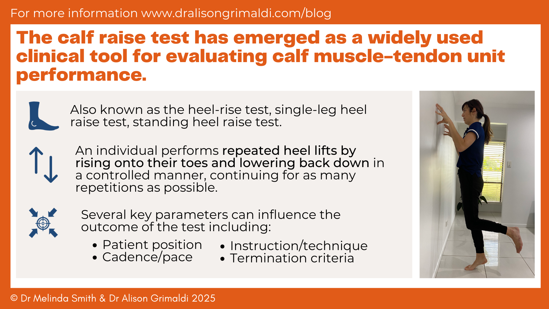
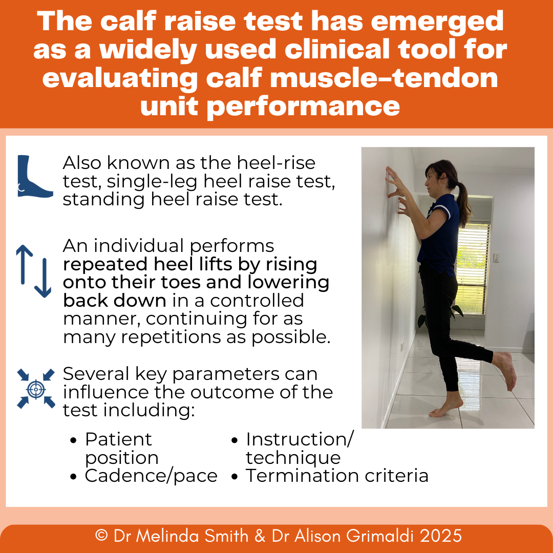
The calf raise test is a clinical tool for assessing the strength-endurance capacity of the calf muscle-tendon unit9, 10 (Figure 3 - below). During this assessment, an individual performs repeated heel lifts by rising onto their toes and lowering back down in a controlled manner, continuing for as many repetitions as possible.
This test challenges the ankle plantar flexors through continuous concentric-eccentric action cycles, providing insight into functional capacity. The calf raise test offers several practical advantages for clinicians, including minimal equipment requirements, quick administration time, limited space needs, and suitability for field-based testing environments. Although commonly performed in unilateral stance it can also be performed in bipedal stance.
Outcome measures from the calf raise test
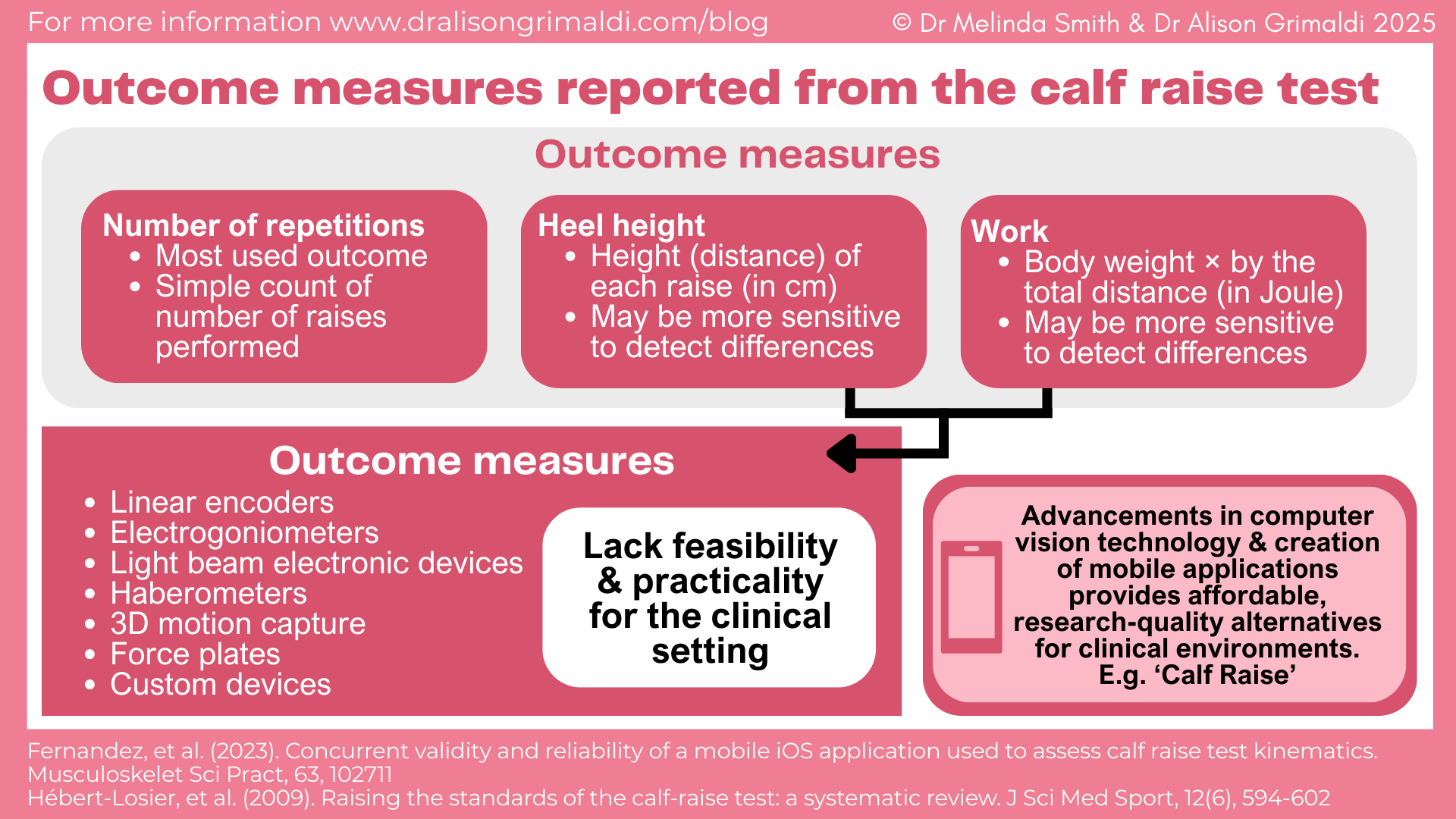
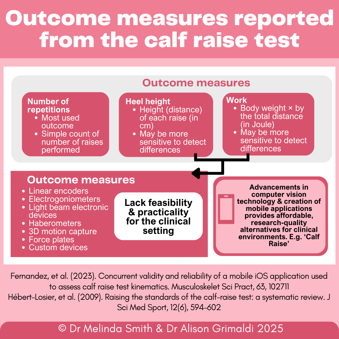
If we look to the literature, outcome measures reported from the calf raise test include number of repetitions, peak height and work11 (Figure 4 - below). Historically, the most reported outcome measurement from the calf raise test is the number of raises performed.9 However other measurements, such as heel lift height, are a critical consideration when assessing calf raise performance as shorter height ranges may lead to increased repetition counts simply because less work is required per repetitions.
‘Work’ accounts for the positive displacement (range) of each repetition and mass of the individual. Consider an individual who performs 20 calf raises on the right leg and 20 calf raises on the left leg, but the left heel lifts to a height of 5 cm compared to 10cm on the right. The amount of work performed on the left is lower than the right. In this instance, simply counting/comparing the number of repetitions may not fully reflect calf capacity. Scientific studies support this as researchers have reported that, in patients with Achilles tendon rupture and Achilles tendinopathy, work is a more sensitive measure for detecting differences than the number of repetitions.10, 12-14
Discover our Foot and Ankle Course
If you enjoyed this blog, you might like to take the online course. With 9.5 hours of online learning content, organised in short, easy-to-digest lessons & modules, explore the different aspects of foot and ankle function and the implications this has for muscle and movement assessment and therapeutic exercise prescription around the foot and ankle.
Factors that influence outcomes of the calf raise test
If we look to the literature, there is substantial variation of key testing parameters that has been described. To ensure reliable and valid results when administering the calf raise test, several methodological aspects should be considered and standardized across testing sessions. These include:
- Patient position
- Cadence
- Instruction/technique
- Test termination criteria
- Individual characteristics
Let’s take a closer look at these factors.
Patient position
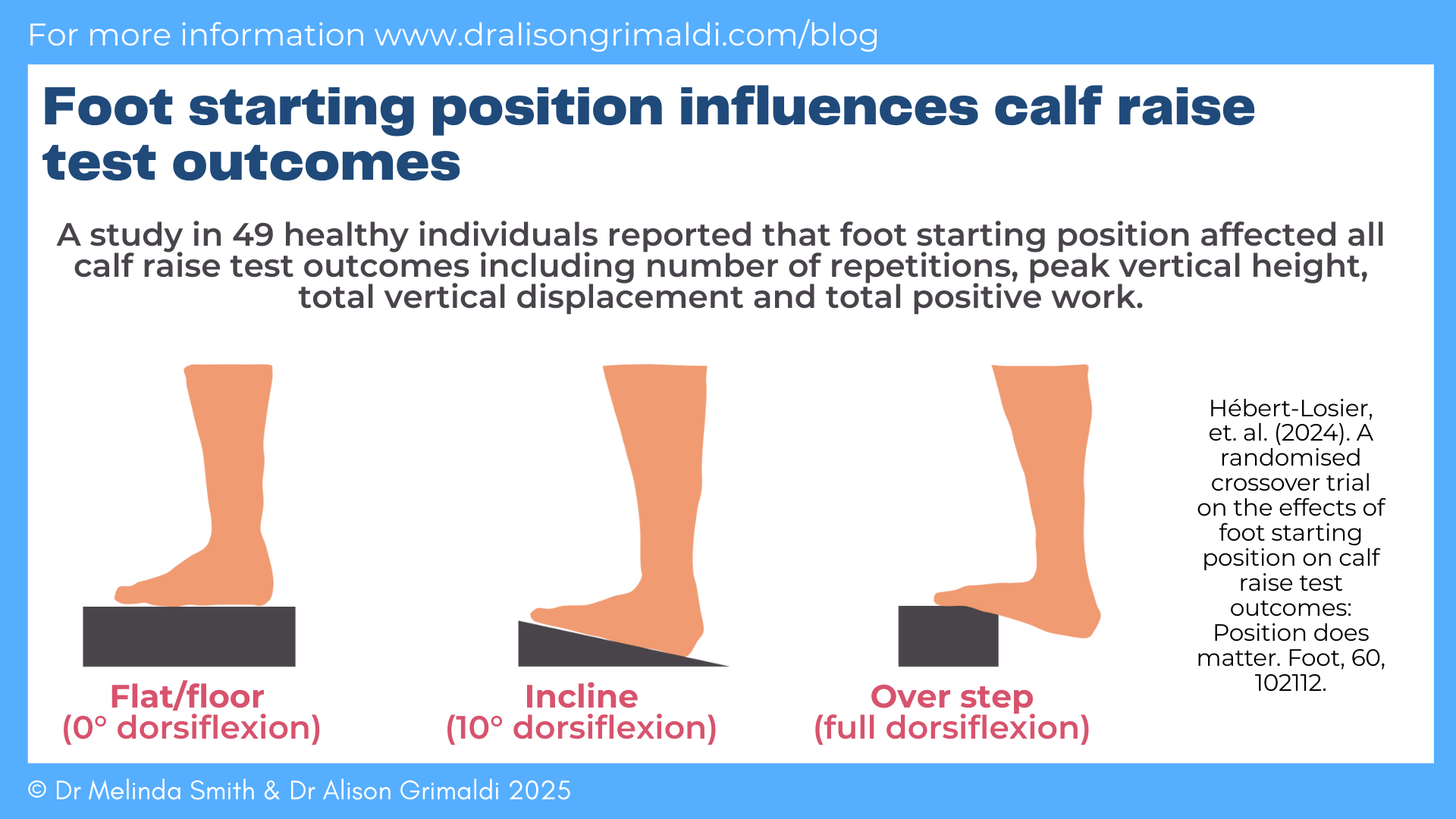
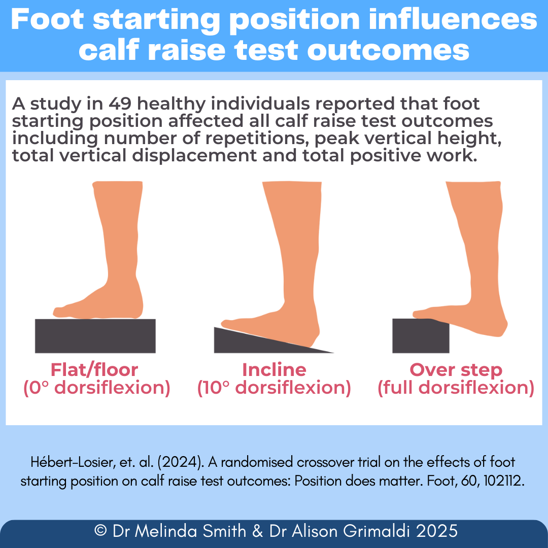
The calf raise test has been described with various starting positions including from plantigrade (from a flat surface or floor), 10 degrees dorsiflexion with an incline platform, and near maximal dorsiflexion with the heel over the edge of a step, see Figure 59. Variation in starting foot position may influence test outcomes. For example, greater mechanical work and earlier calf muscle fatigue may occur due the increased range of motion from starting positions that are more dorsiflexed. This could lead to a fewer number of repetitions being performed. Or, it is also possible that due to the length-tension relationship, starting in a position of greater dorsiflexion range may produce greater plantarflexion force and result in a higher number of repetitions being performed. Hébert-Losier and colleagues16 investigated the effect of these three starting foot positions on calf raise test outcomes in forty-nine healthy individuals. They reported that foot starting position affected all calf raise test outcomes including number of repetitions, peak vertical height, total vertical displacement and total positive work. Performing the calf raise test from the flat floor produced 23% and 39% more repetitions compared to a 10-degree incline or with the heel over the edge of a step.
Knee joint position is another parameter of the calf raise test that is often manipulated in the clinical setting. Based on anatomic and physiologic principles, knee position has traditionally been used to influence triceps surae selectivity - an extended knee position for the gastrocnemius muscles and a flexed knee (45 degrees) for the soleus muscle. In forty-eight healthy volunteers, Hébert-Losier and colleagues17 investigated the influence of knee joint angle on triceps surae muscle fatigue during heel raises performed to volitional fatigue. All three muscles fatigued during the testing with greater fatigue in the gastrocnemius muscles compared to the soleus. This is perhaps not surprising given muscle physiology and fibre type differences among the triceps surae muscles. What was perhaps surprising was that knee joint position (extended or 45 degrees flexion) did not influence muscle fatigue. Gastrocnemius and soleus fatigue was similar for the calf raise test performed with the knee extended and flexed. These findings challenge our traditional thoughts on triceps surae selectivity and suggest that either knee joint position is useful in assessing any one of the triceps surae muscles.
Cadence
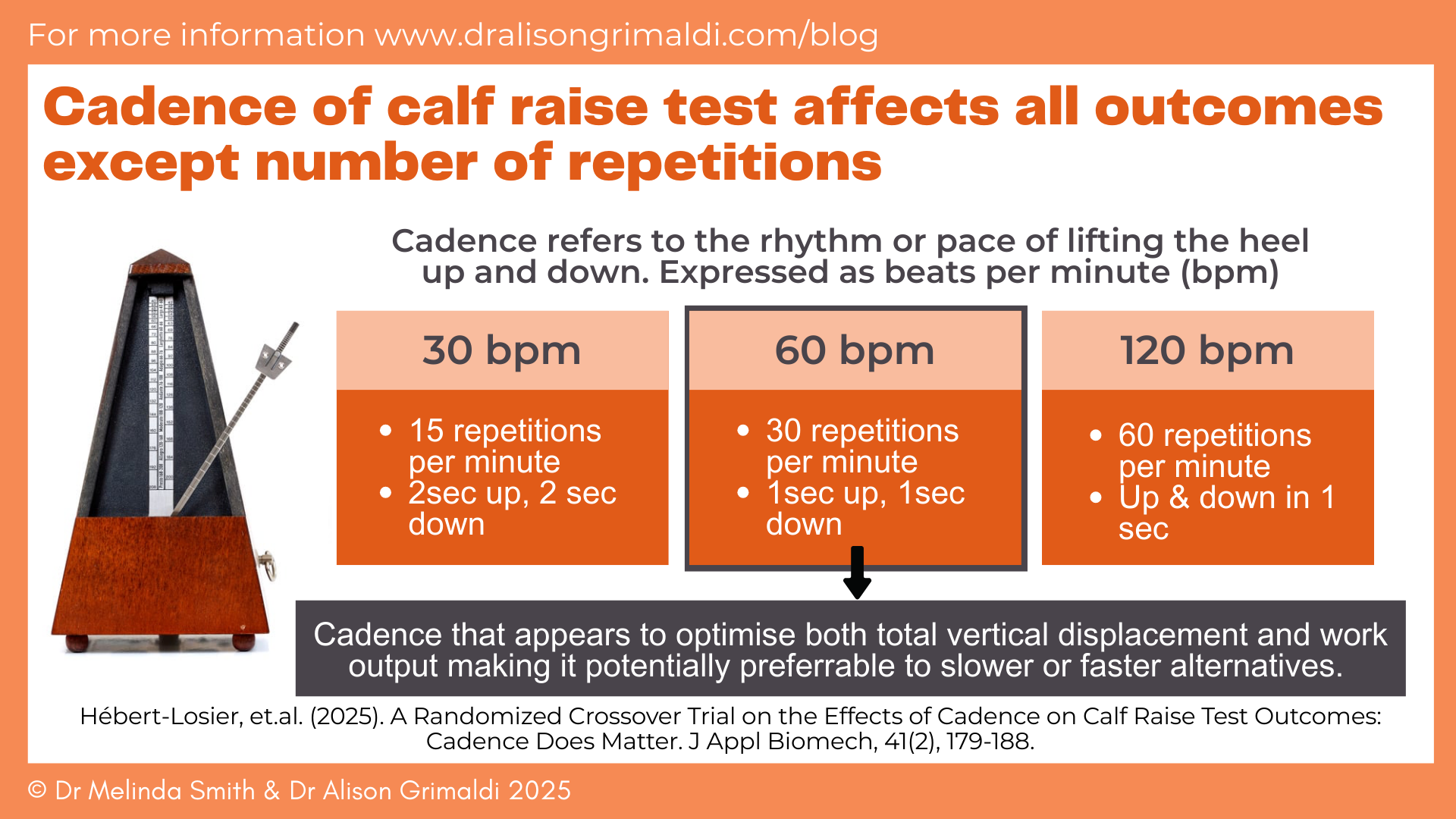
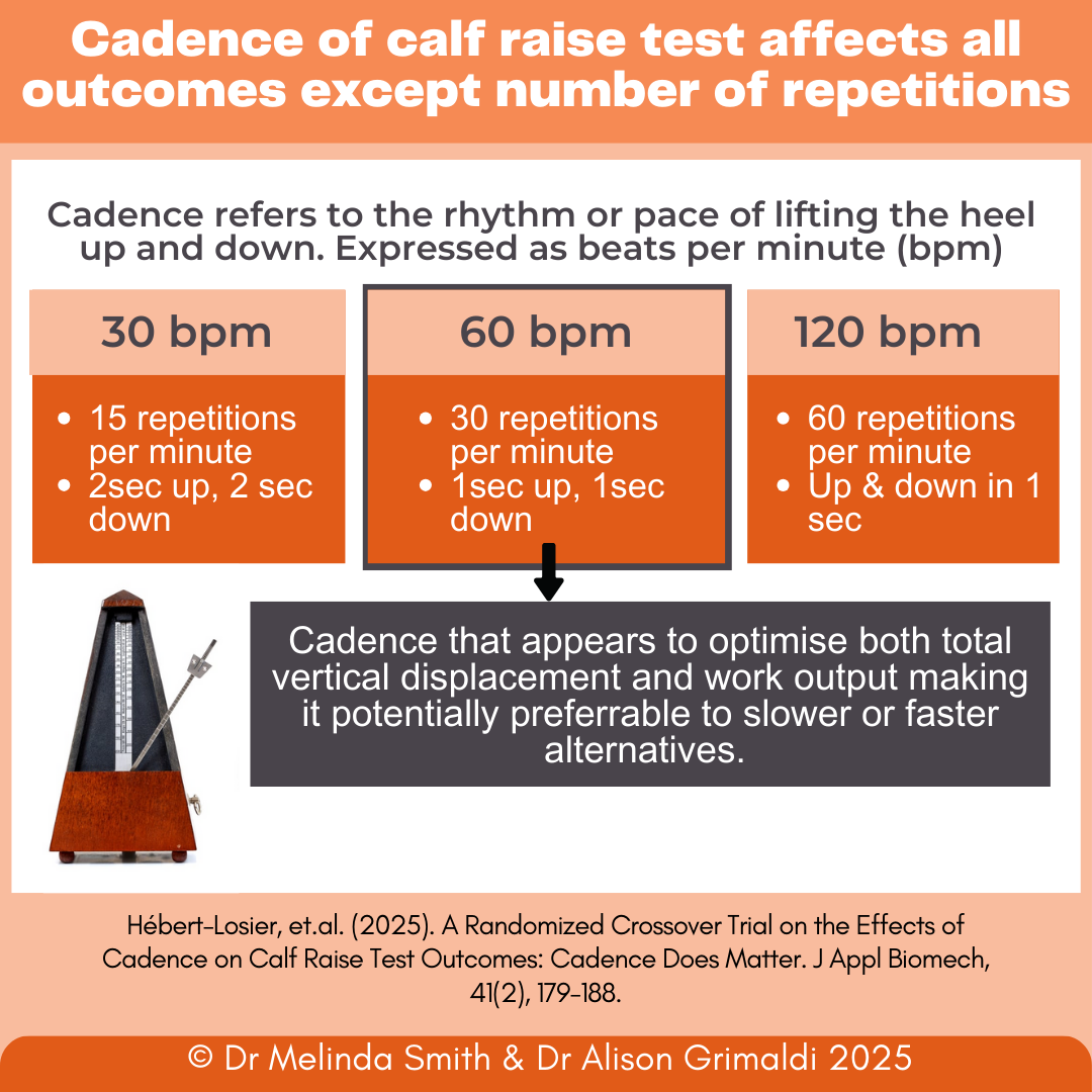
Cadence is used to describe the pace or regular rhythm at which a task is performed, for example, in walking it is the number of steps taken per minute. In the context of the calf raise test cadence refers to the timing of the heel lifting up and down. For example, a cadence of 60 beats per minute would result in 30 calf raise repetitions per minute with an individual lifting their heel up on one beat (one second) and down in one beat (one second). A metronome is often used to control cadence during the calf raise test. A variety of difference cadences have been reported from 30 to 120 beats per minute9, 18. The cadence of the test may influence the outcome because movement velocity can influence musculotendinous unit mechanics19. In a study of 36 healthy individuals, Hébert-Losier and colleagues 20 examined the effect of three cadences (30, 60 and 120 beats per minute) on calf raise test outcomes, Figure 6. They reported that cadence influenced all outcomes (total vertical displacement, total work, peak height, peak power) except the number of repetitions. The authors concluded that a moderate cadence of 60 beats per minute appears to optimise both total vertical displacement and work output making it potentially preferrable to slower or faster alternatives. However, the ideal cadence may ultimately depend on specific training objectives that relate to the assessment. The key message is whichever cadence you decide to use make sure that you control it (such as using a metronome) to ensure consistency between assessments, particularly when outcomes other than number of repetitions are of interest.
Instructions, technique & test termination criteria
In addition to foot/knee position and cadence, other parameters may also influence test outcomes. In fifty-one Australian football players, Green and colleagues 21 compared the results of cued and non-cued calf raise tests. In the non-cued version of the test individuals were barefoot, faced a wall with fingertip support at shoulder level and were simply told to lift their heel up and down as many repetitions as possible i.e. the test was performed at a self-selected pace and ceased based on volitional failure. In the cued version of the test individuals were given additional instruction on how to perform the test and the test ceased based on either volitional or technical failure. Additional instructions included to keep pace with a metronome at 60 beats per minute, perform maximum calf raise height with each repetition, keep the knee straight, align the middle of the ankle over the second toe, and no rocking of the body back and forth. The test was ceased due to technical failure if for two consecutive repetitions, with a warning provided, the individual was unable to maintain metronome pace or were unable to maintain technical cues resulting in knee flexion, forward trunk lean or rocking, hip strategy, reduced heel height or foot and ankle alignment/position error. During the cued calf raise test individuals performed on average 12 less repetitions than the non-cued test. These findings demonstrate that how the test is performed influences the number of repetitions achieved and highlights the importance of standardised test parameters.
Other test parameters to considerations
There are several other aspects of executing the calf raise test that have been described among studies that feasibly could influence outcomes, but have not been well explored in the literature. These include:
- providing standard encouragement during the test such as “keep going as high as possible”
- Performing the test barefoot or in shoes
- Provide a warm-up & familiarisation/practice. e.g. a warm-up of 10 bilateral calf raises followed by a familiarisation/practice of 3 single-leg calf raises as per instructions and with metronome pace.
When using the calf raise test clinicians should consider all of these elements that may influence the outcome (Figure 7), and select a standardised approach.
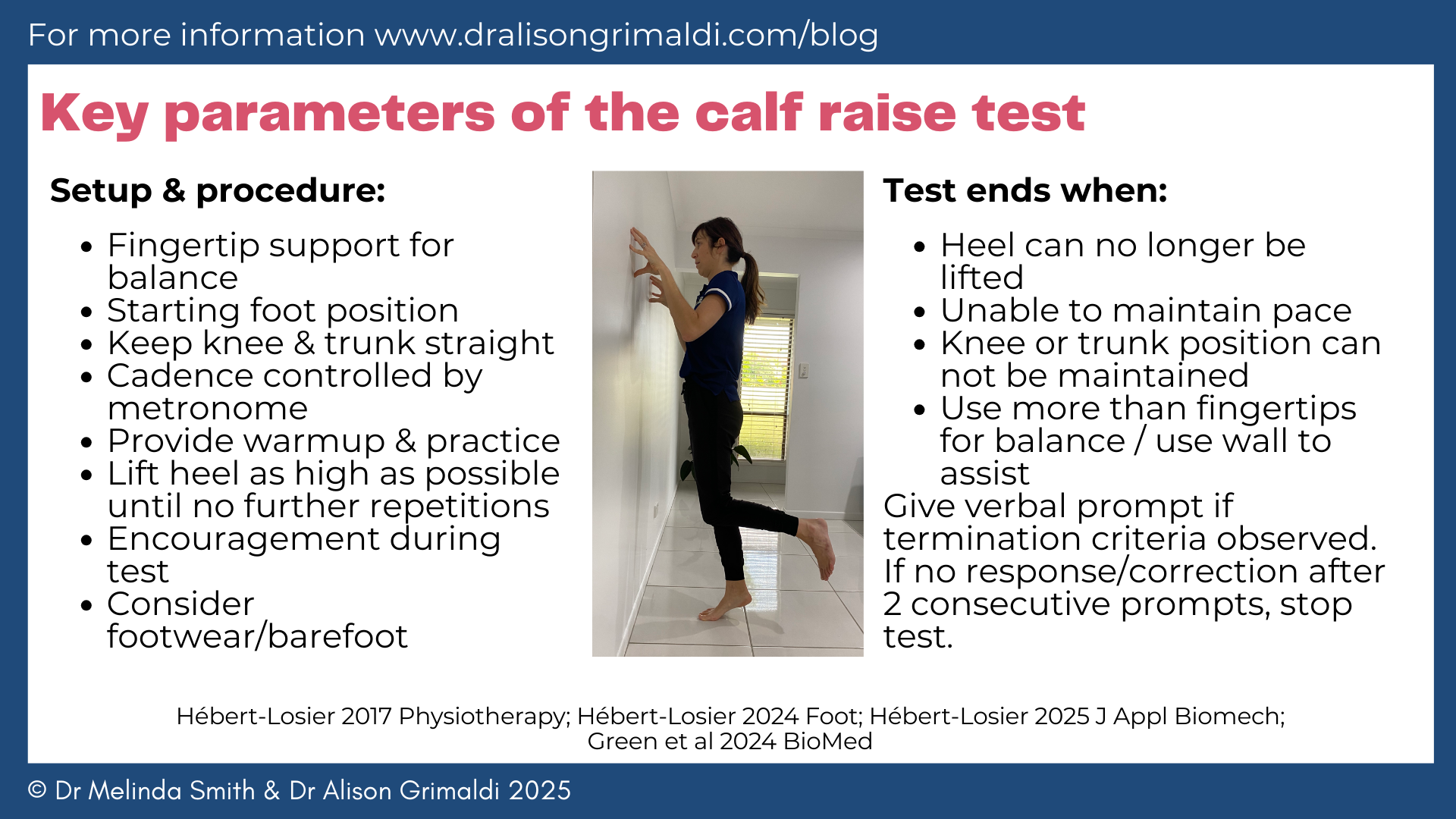
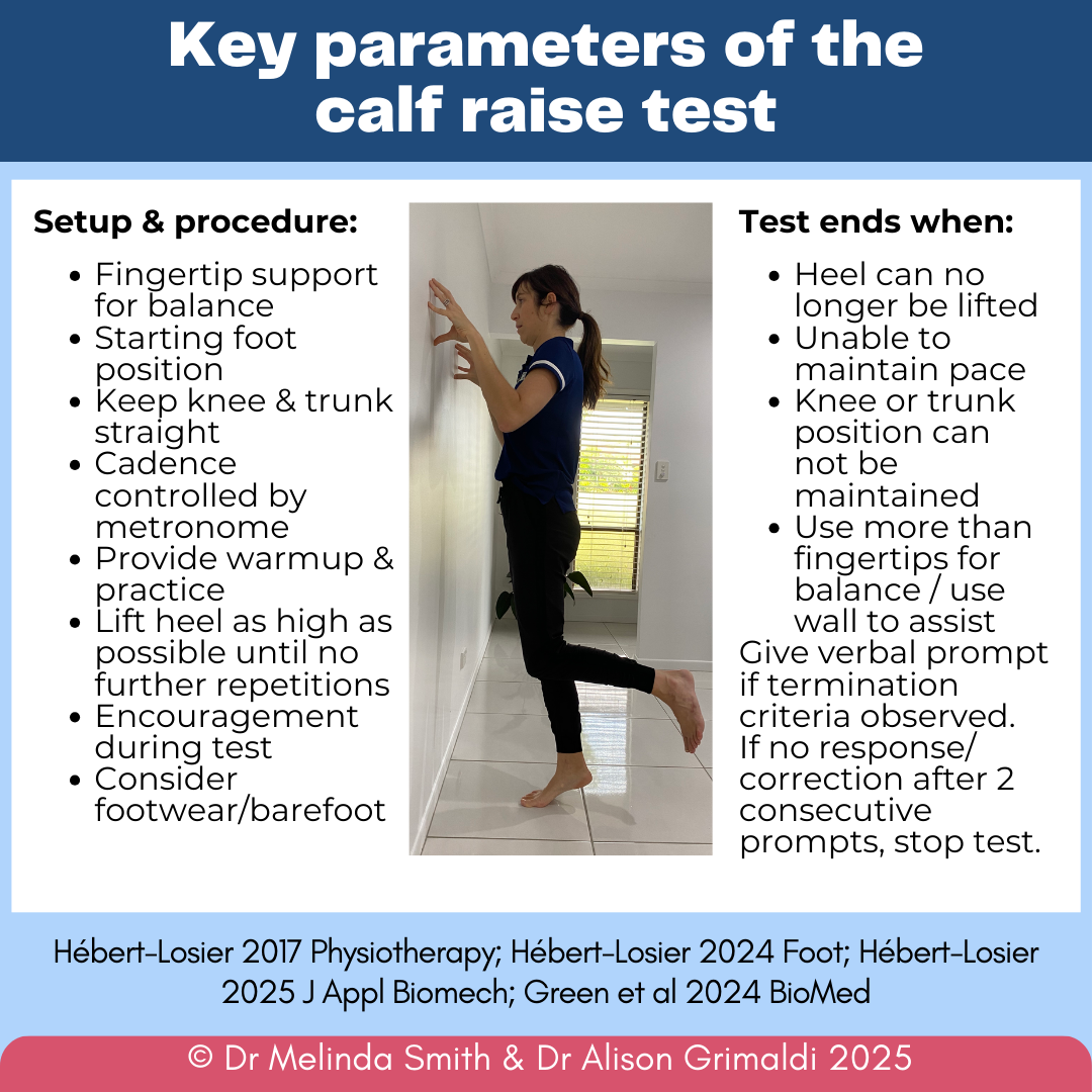
Several key parameters influence outcomes of the calf raise test and therefore using a standardised protocol is important to ensure reliable and valid outcomes.
Individual characteristics
Research studies have reported personal characteristics that are associated with outcomes of the calf raise test. From a cohort of 500 healthy adults, Visser and colleagues22reported that female sex, lower body mass index and lower physical activity levels were associated with lower calf raise test results. This was like a previous study in 566 healthy adults that reported female sex, older age and lower physical activity level had a negative effect of calf raise test outcome23. Research has reported similar findings in other populations. Green and colleagues21 reported in 51 Australian football athletes that female sex, age over thirty years, and indigenous ethnicity were associated with reduced calf raise test repetitions. In 165 children aged 10-17 years, children who were older, had lower body mass index and were more active showed superior calf raise test outcomes24. Studies also seem to suggest that calf raise test outcomes are not different between limbs including left/right and dominant/non-dominant comparisons22-24.
Interpretation of calf raise test outcomes
Normative data
To accurately interpret calf raise test results it is important to remember factors that may influence outcomes (i.e. key test parameters and personal characteristics discussed above). Normative data has been reported for various populations18 including healthy adults22, 23, children24 and athletes25. To most accurately reference normative data, it is important to ensure that the calf raise test is performed that same was as the normative data describes (because key parameters such as start position, cadence, instructions, termination criteria can influence results). Given the influence of personal characteristics, studies often provide normative data by sex and age.
One group of researchers has provided a web-based tool for clinicians to estimate normative values for calf muscle strength-endurance assessed using the calf raise test via the Calf Raise application (www.achillestendontool.com/HRET)22. This involves inputting the individual’s sex, age, weight and height (body mass index is calculated) and physical activity level to generate estimates for repetitions, peak height and work (as well as vertical displacement and peak power). It is interesting that from studies in healthy adults and children (10-17 years), researchers report that the median number of repetitions is 25, which aligns to the historical criterion for normal recommended by Lunsford and Perry26.
Determining change
The calf raise test has demonstrated high test-retest reliability23, 27. Studies report a standard error of measurement of two repetitions and minimal detectable change of six repetitions. Applied to the clinical setting, this means six repetitions are regarded as the minimal amount of change that needs to be observed (e.g. between two assessments/time-points) for it to be considered a real change.
When to use the calf raise test
Given the important contribution of the calf muscles to lower limb function, assessing calf muscle capacity via the calf raise test is of interest in many athletic and clinical populations. For example, people with:
- Achilles tendinopathy
- Achilles tendon rupture
- Calf muscle injury
- Plantar heel pain
- Medial tibial stress syndrome
- Tendinopathies of accessory plantar flexors such as flexor hallucis longus, tibialis posterior
Or, anyone who walks, runs or jumps!
In the literature the calf raise test has been described in clinical populations across several disciplines including people with Achilles tendinopathy28, Achilles tendon rupture13, 14, plantar heel pain29, peripheral arterial disease30, upper motor neuron lesion due to stroke31, children24, 32 and older adults33.
The calf raise test can be used to evaluate impairments, monitor recovery/progress and evaluate effects of rehabilitation. Remember, when comparing over time/between sessions, 6 repetitions is considered a real change.
Calf raise test outcomes have been associated with functional performance in several populations including 10m sprint performances in rugby players34, walking speed & ankle power in surgically treatment Achilles tendon rupture35, locomotor capacity in children, functional fitness in older adults33 and functional capacity in people with peripheral arterial disease30.
Summary of key point
As the scientific evidence has demonstrated, several key parameters influence outcomes of the calf raise test and therefore using a standardised protocol is important to ensure reliable and valid outcomes. Although no standardised protocol has been published and widely accepted, clinicians should consider standardising key elements that have been shown to influence the outcome such as patient position, cadence, technique and termination criteria. Other considerations when executing the test in the clinical setting include providing a warm-up and practice prior to the test, standard encouragement during the test and use of footwear or barefoot. Finally, it is important to understand that individual characteristics also influence calf raise test outcomes including age, sex, body mass index, ethnicity and physical activity level.
We hope you’ve found this blog on the calf raise test helpful.
Want to help more people who visit your clinic
with foot & ankle pain?
To learn more about integrating foot and ankle muscle assessment and therapeutic exercise into the clinical setting take a look at our Mastering Movement of the Foot and Ankle online course or practical workshop.
Check Out Some More Foot and Ankle Blogs:
References
- Standring S. Gray's Anatomy: The Anatomical Basis of Clinical Practice. Forty-second edition edn. New York: Elsevier; 2021.
- Bolsterlee B, Finni T, D'Souza A, Eguchi J-i, Clarke EC, Herbert RDJP. Three-dimensional architecture of the whole human soleus muscle in vivo. 2018; 6.
- Roberts TJ. The integrated function of muscles and tendons during locomotion. Comparative Biochemistry and Physiology Part A: Molecular & Integrative Physiology 2002; 133(4):1087-1099.
- Perry J, Burnfield JM. Gait Analysis: Normal and Pathological Function. Second edition edn. Milton: CRC Press; 2010.
- Sadeghi H, Sadeghi S, Prince F, Allard P, Labelle H, Vaughan CL. Functional roles of ankle and hip sagittal muscle moments in able-bodied gait. Clinical biomechanics (Bristol) 2001; 16(8):688-695.
- Lichtwark GA, Bougoulias K, Wilson AM. Muscle fascicle and series elastic element length changes along the length of the human gastrocnemius during walking and running. Journal of Biomechanics 2007; 40(1):157-164
- Hamner SR, Seth A, Delp SL. Muscle contributions to propulsion and support during running. J Biomech 2010; 43(14):2709-2716.
- Dorn TW, Schache AG, Pandy MG. Muscular strategy shift in human running: dependence of running speed on hip and ankle muscle performance. The Journal of experimental biology 2012; 215(Pt 11):1944-1956.
- Hébert-Losier K, Newsham-West RJ, Schneiders AG, Sullivan SJ. Raising the standards of the calf-raise test: a systematic review. Journal of science and medicine in sport 2009; 12(6):594-602.
- Fernandez MR, Hébert-Losier K. Devices to measure calf raise test outcomes: A narrative review. Physiother Res Int 2023; 28(4):e2039.
- Fernandez MR, Athens J, Balsalobre-Fernandez C, Kubo M, Hébert-Losier K. Concurrent validity and reliability of a mobile iOS application used to assess calf raise test kinematics. Musculoskelet Sci Pract 2023; 63:102711.
- Silbernagel KG, Gustavsson A, Thomeé R, Karlsson J. Evaluation of lower leg function in patients with Achilles tendinopathy. Knee Surg Sports Traumatol Arthrosc 2006; 14(11):1207-1217.
- Silbernagel KG, Nilsson-Helander K, Thomeé R, Eriksson BI, Karlsson J. A new measurement of heel-rise endurance with the ability to detect functional deficits in patients with Achilles tendon rupture. Knee Surg Sports Traumatol Arthrosc 2010; 18(2):258-264.
- Silbernagel KG, Steele R, Manal K. Deficits in Heel-Rise Height and Achilles Tendon Elongation Occur in Patients Recovering From an Achilles Tendon Rupture. Am J Sports Med 2012; 40(7):1564-1571.
- Ashnai F, Lindskog J, Brorsson A, Nilsson-Helander K, Beischer S. The Calf Raise App shows good concurrent validity compared with a linear encoder in measuring total concentric work. Knee Surg Sports Traumatol Arthrosc 2024; 32(8):2170-2177.
- Hébert-Losier K, Fernandez MR, Athens J, Kubo M, O'Neill S. A randomised crossover trial on the effects of foot starting position on calf raise test outcomes: Position does matter. Foot (Edinb) 2024; 60:102112.
- Hébert-Losier K, Schneiders AG, García JA, Sullivan SJ, Simoneau GG. Influence of Knee Flexion Angle and Age on Triceps Surae Muscle Fatigue During Heel Raises. J Strength Cond Res 2012; 26(11):3134-3147.
- Bohannon R, W. . The heel-raise test for ankle plantarflexor strength : a scoping review and meta-analysis of studies providing norms. Journal of Physical Therapy Science 2022; 34(7):528-531.
- Alcazar J, Csapo R, Ara I, Alegre LM. On the Shape of the Force-Velocity Relationship in Skeletal Muscles: The Linear, the Hyperbolic, and the Double-Hyperbolic. Front Physiol 2019; 10:769-769.
- Hébert-Losier K, Fernandez MR, Athens J, Kubo M, O'Neill S. A Randomized Crossover Trial on the Effects of Cadence on Calf Raise Test Outcomes: Cadence Does Matter. J Appl Biomech 2025; 41(2):179-188.
- Green B, Coventry M, Pizzari T, Rio EK, Murphy MC. Form Matters—Technical Cues in the Single Leg Heel Raise to Failure Test Significantly Change the Outcome: A Study of Convergent Validity in Australian Football Players. 2024. Accessed:
- Visser TSS, Neill SO, Hébert-Losier K, Eygendaal D, de Vos RJ. Normative values for calf muscle strength-endurance in the general population assessed with the Calf Raise Application: A large international cross-sectional study. Braz J Phys Ther 2025; 29(3):101188.
- Hébert-Losier K, Wessman C, Alricsson M, Svantesson U. Updated reliability and normative values for the standing heel-rise test in healthy adults. Physiotherapy 2017; 103(4):446-452.
- Hébert-Losier K, Pandit Y, Wilson OWA, Clarke J. Looking Beyond the Number of Repetitions: An Observational Cross-Sectional Study on Calf Raise Test Outcomes in Children Aged 10-17 Years. Phys Occup Ther Pediatr 2025; 45(2):240-255.
- Hébert-Losier K, Ngawhika TM, Gill N, Balsalobre-Fernandez C. Validity, reliability, and normative data on calf muscle function in rugby union players from the Calf Raise application. Sports Biomech 2022; ahead-of-print(ahead-of-print):1-22.
- Lunsford BR, Perry J. The Standing Heel-Rise Test for Ankle Plantar Flexion: Criterion for Normal. Phys Ther 1995; 75(8):694-698.
- Ross MD, Fontenot EG. Test–Retest Reliability of the Standing Heel-Rise Test. Journal of sport rehabilitation 2000; 9(2):117-123.
- Murphy M, Rio E, Debenham J, Docking S, Travers M, Gibson W. Evaluating the progress of mid-portion achilles tendinopathy during rehabilitation: a review of outcome measures for muscle structure and function, tendon structure, and neural and pain associated mechanisms. Int J Sports Phys Ther 2018; 13(3):537-551.
- Sullivan J, Burns J, Adams R, Pappas E, Crosbie J. Musculoskeletal and Activity-Related Factors Associated With Plantar Heel Pain. Foot Ankle Int 2015; 36(1):37-45.
- Oliveira AKMLM, Neves VRP, Trevizan PFP, Oliveira RDBPT, Pereira DAGP. Heel Rise Test Accuracy in the Assessment of the Functional Capacity of Individuals With Peripheral Arterial Disease. J Manipulative Physiol Ther 2024; 47(5-9):107-113.
- Svantesson U, Osterberg U, Grimby G, Sunnerhagen KS. The standing heel-rise test in patients with upper motor neuron lesion due to stroke. Scand J Rehabil Med 1998; 30(2):73-80.
- Maurer C, Finley A, Martel J, Ulewicz C, Larson CA. Ankle Plantarflexor Strength and Endurance in 7-9 Year Old Children as Measured by the Standing Single Leg Heel-Rise Test. Physical & occupational therapy in pediatrics 2007; 27(3):37-54.
- André H-I, Carnide F, Moço A, Valamatos M-J, Ramalho F, Santos-Rocha R, Veloso A. Can the calf-raise senior test predict functional fitness in elderly people? A validation study using electromyography, kinematics and strength tests. Phys Ther Sport 2018; 32:252-259.
- Hébert-Losier K, Ngawhika TM, Balsalobre-Fernandez C, O'Neill S. Calf muscle abilities are related to sprint performance in male Rugby Union players. Physical therapy in sport 2023; 64:117-122.
- Nordenholm A, Hamrin Senorski E, Nilsson Helander K, Möller M, Zügner R. Greater heel-rise endurance is related to better gait biomechanics in patients surgically treated for chronic Achilles tendon rupture. Knee Surg Sports Traumatol Arthrosc 2022; 30(11):3898-3906.

