Anterior Hip Pain: Causes and contributing factors

Anterior hip pain is one of the most common presentations of hip pain we see in clinical practice as health professionals. Despite its regular presentation, all cases are certainly not the same! There are quite a range of different potential sources of nociception or referral, contributing factors and associated impairments. Adequate consideration of anterior hip pain causes and contributing factors is important for optimal outcomes of interventions.
A lack of appreciation for anterior hip pain causes and contributing factors may lead to generic application of a narrow range of clinical tests and treatment techniques, which often fails to adequately provide the answers and the relief our patients need. In this blog, I’m going to bring together some key information on this topic and signpost where you can find more detail.
These are the topics we’ll be covering in this blog:
What causes anterior hip pain:
Joint Related Anterior Hip Pain
Soft tissue related anterior hip pain
Nerve related anterior hip pain
Factors that may contribute to the development of anterior hip pain and pathology:
Discover our Anterior Hip & Groin Pain Course
If you enjoyed this blog, you might like to take the online course on Anterior Hip & Groin Pain - 5 hours of guided online video content. Better your skills and understanding of the anterior hip and groin and become equipped with the knowledge to administer clinical diagnostic tests and management strategies.
What causes anterior hip pain?
Causes of pain are still a bit of an enigma, but thanks to pain scientists we now understand that pain is an output of the brain, an experience generated from a wide range of inputs. Central processing and psychosocial contributors are key considerations and there are plenty of great resources available that cover these topics.
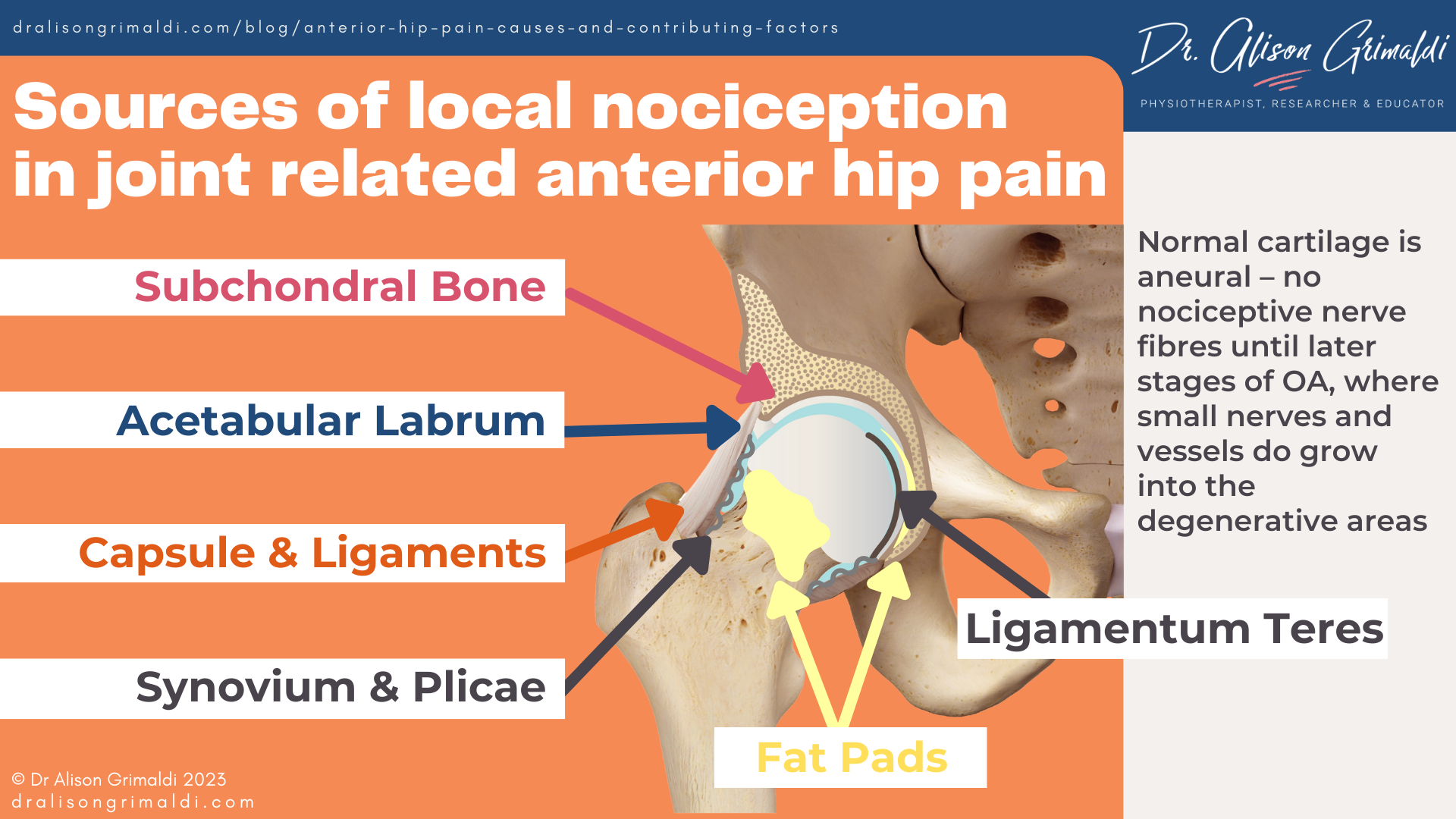
For our purposes in this blog, we’ll be focusing on local nociceptive sources of anterior hip pain. Local nociception remains an important contributor to most mechanical musculoskeletal pain presentations.
So, what sources of nociception exist at the anterior hip, and does it really matter where the pain is coming from? Let’s address sources of nociception first. I find it helpful to use a framework to think through the possible sources of local nociception, based around joint related pain, soft tissue related pain, nerve related pain and bone related pain.
The relevance of intra-articular hip pathology to pain
There is plenty of scientific information now available that highlights the mismatch between the presence of pathology and the role that pathology plays in an individual’s pain presentation, but they are closer associations with some types of pathology than others.
Acetabular labral tears
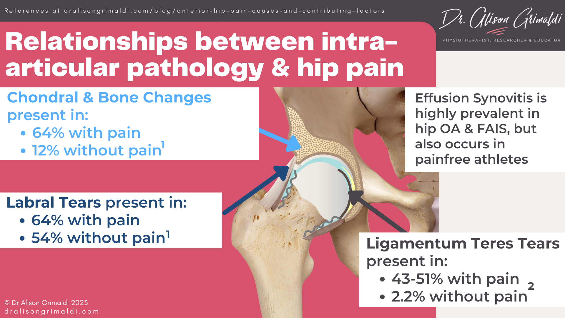
In 2018, a systematic review and meta-analysis reported that while 64% of those with hip pain had labral tears, 54% of those without pain also had labral tears on imaging.1 This means that while labral tears can certainly contribute to anterior hip pain, just having a tear doesn’t mean that they are causing that pain. Thus, treatment should not be determined by imaging findings.
However, there are important functions of the acetabular labrum, and pathology may have relevance to joint health even if this pathology is not directly related to the pain experience.
Read more about the functions of the labrum and impact of pathology on joint health by clicking on the pink text.
Chondral and bone changes
Heerey’s 2018 review reported that while 64% of those with hip pain had cartilage defects, only 12% of those without pain also had defects on imaging.1 However, we’ve already mentioned above that cartilage is probably not a primary source of nociception until later stages of hip osteoarthritis.
From these statistics, there would seem to be a potential relationship between chondral pathology and hip pain, but this may be mediated by adjacent nociceptive structures that may be impacted by deterioration in cartilage health - for example the synovium or subchondral bone.
Bone marrow lesions appear much more likely to occur in those with painful hips, although the database available is not yet large enough to make firm conclusions. Subchondral bone is certainly innervated and may be a source of nociception, and there is an important interplay between subchondral bone and the overlying cartilage with respect to maintaining joint health.
Read more about the relevance and impact on chondral pathology and bone changes on hip pain and joint health by clicking on the pink text.
Ligamentum teres tear
Information available for prevalence of ligamentum teres tears, shows clearly that lig teres tears are far more common in those with hip symptoms: while 43-51% of those with hip pain had ligamentum teres pathology, only 2.2% of those without pain displayed pathology.2
In recent years there has also been much more attention paid to this important ligament with respect to its relationship with joint health and outcomes of intervention for joint related hip pain.
Ligamentum teres injury has been linked with:
- High prevalence of labral and chondral pathology – femoral head and acetabular cartilage defects.2
- Higher rates of failure of non-surgical management – those with lig teres tear and a labral tear have been shown to have a 16.2 times greater risk of surgery than those with labral tear alone.3
- Higher rates of failure of surgical intervention and progression to total hip arthroplasty after arthroscopy for Femoroacetabular Impingement Syndrome (FAIS)4 or dysplasia.5
And there is still much to learn about this ligament.
Read more about the relevance of the ligamentum teres by clicking on the pink text.
Effusion Synovitis
Effusion synovitis is highly prevalent in those with painful hip osteoarthritis and cam FAIS, but athletes without symptoms may also display effusion, presumably a response to loading. The relationship between effusion and pain is not yet clear, but the synovium is highly innervated and certainly a potential source of nociception.
Effusion synovitis has also been implicated in the pathogenesis and progression of hip osteoarthritis, suggesting it is not something to be ignored.
Read more about the relevance of effusion synovitis by clicking on the pink text.
Does it matter if the pain is in the joint or the soft tissues?
Well, it depends on the treatment approach. If the approach to soft tissue related anterior hip pain is stretching, massage, needling and even corticosteroid injections, these interventions may not only be ineffective in the longer term, but may be provocative for the joint, hindering optimal progress.
If the approach to treatment involves a close assessment of factors that might be overloading the anterior hip region, many features of a load management and exercise program for joint related pain and soft tissue related pain are likely to be consistent, and helpful for both.
Factors that may contribute to the development of anterior hip pain and pathology
Identifying key underlying drivers for anterior hip pain may be very useful in the development of an optimally effective intervention. Multiple factors are often at play, with varying combinations relevant for different individuals.
There will usually be a combination of non-modifiable factors such as genetics, sex, past traumatic events and bony morphology (well, non-modifiable by non-surgical health professionals), and modifiable factors such as muscle and movement impairments, and load profile (type and volume of activity etc). Psychosocial contributors will play larger roles for some than others but should always be considered.
While modifiable contributing factors for anterior hip pain might appear the most logical to identify and address, awareness of non-modifiable factors can also be extremely important in managing physical loads in the region and developing the most helpful education and exercise strategies.
Natural collagen structure and extensibility may play a role in the development of hip pain and degenerative change. Those with Ehlers Danlos Syndrome or Hypermobile Spectrum Disorders may require some variations in treatment approach for anterior hip pain.
The information below will focus on the potential roles of bony morphology, muscle, movement patterns and joint loading on articular health and joint related anterior hip pain.
Bone morphology, hip pain and pathology
The relevance of bone morphology in those with hip pain is often debated. For some, it plays only a minor role or no role at all. For others with more severe bony variations, morphology might be the primary factor underpinning the development of anterior hip pain, or there may be key interactions between morphology and movement or muscle impairments that amplify the impact of that morphology.
It is always worth screening for potential morphological factors at the beginning of any patient’s rehabilitation journey, rather than discovering them months or years later, after many unsuccessful or only partly successful attempts at rehabilitation.
Cam morphology and Femoroacetabular Impingement Syndrome
What is cam morphology?
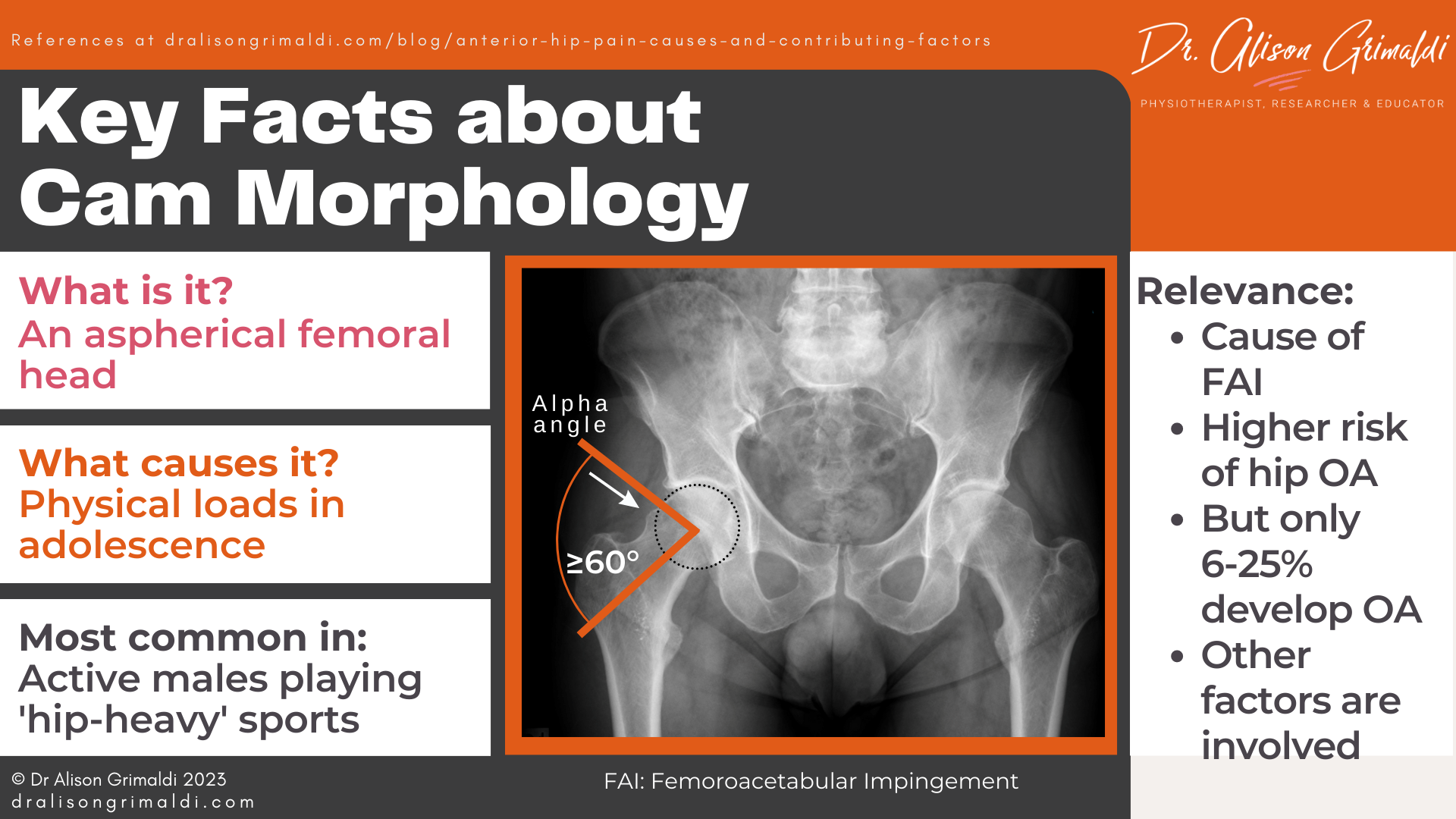
Cam morphology is an aspherical femoral head, where extra bone at the femoral head-neck junction may cause early impingement of the femoral head-neck junction and the acetabulum during end-range hip motion – referred to as Femoroacetabular Impingement (FAI).
The most common definition of cam morphology uses an alpha angle threshold of 60° on imaging. The alpha angle is the angle between a line from the centre of the femoral head through the middle of the femoral neck and a line that crosses the point at which the bone of the head of the femur leaves the circular constraints of a typical femoral head (see the graphic below). The higher the angle, the more severe the cam morphology (larger bump at the head-neck junction).
What causes cam morphology?
Cam morphology develops primarily during adolescence, when the growth plate is open.
Development of cam morphology appears to be related to:
- Sex – more common in males,
- Activity levels – more common in active individuals, and
- Type of sport – more common in ‘hip heavy’ sports.
It is thought that certain physical loads placed across the capital femoral epiphysis may change the orientation of the lateral aspect of the growth plate and cause asymmetric closure of the growth plate, changing the shape of the femoral head and neck.
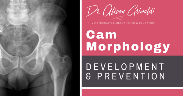
Read more about cam morphology development and prevention by clicking on the pink text.
What is the clinical relevance of cam morphology?
What does it matter? For the majority of people, cam morphology is unlikely to ever cause an issue, but for others with more severe cam morphology and/or those who regularly force their hip into a position of impingement, hip pain and osteoarthritis (OA) may develop over time.
Key facts about cam morphology and hip osteoarthritis:
- 6 - 25% of people with cam morphology will develop hip OA within 5 -19 years.6
- Those with a pathological cam (>78° alpha angle) have increased risk of advanced OA, especially if their hip internal rotation is less than 20°.7
- Those with an alpha angle greater than 85° have a 33% chance of developing OA in next 5 years.7
- Those with cam morphology have a 3-fold great risk of undergoing a total hip replacement over a 14-year period.8
Please note, these studies have been conducted on people who are generally older than 45 years. Further large prospective studies are required to clarify the risks in earlier life.
Read more about The Prevalence of Cam Morphology and its Association with Development of Hip Osteoarthritis by clicking on the pink text.
Other studies are furthering our understanding on bone shape and hip OA risk through 3-D modelling of the proximal femur and acetabulum.
Read more about this paper on bone shape and hip osteoarthritis by clicking on the pink text.
The relationship with cam morphology and hip pain (Femoroacetabular Impingement Syndrome) is less clear and may depend on various other activity/task-specific and general health factors. A recent prospective study found a link with hip pain and cam morphology in those with higher Body Mass Index.8
Acetabular dysplasia
What is acetabular dysplasia?
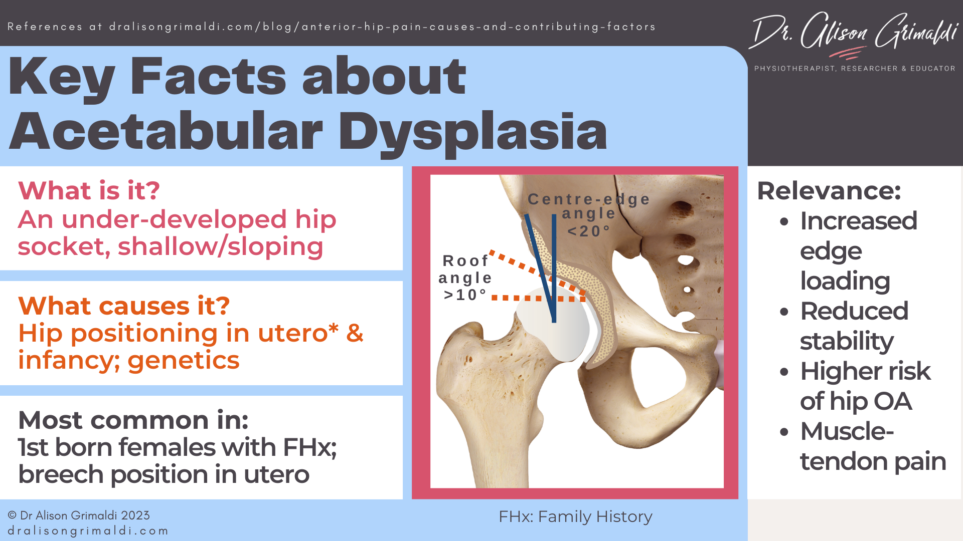
Acetabular dysplasia refers to abnormal or under-development of the hip socket. The traditional model of acetabular dysplasia describes 2 main types of dysplasia: i) a shallow, upward sloped socket and ii) a socket with a short roof – visualized laterally on an AP X-Ray.
However, this definition underappreciates other forms of dysplasia that might result in focal rather than global undercoverage of the femoral head. Wilkin and colleagues proposed a contemporary definition that redefined acetabular dysplasia as a 3-dimensional bony deficiency that considers focal anterior and posterior undercoverage of the femoral head by the acetabulum, as well as the more commonly recognized global undercoverage.9
Read more here to ensure you are seeing the whole picture when it comes to acetabular dysplasia .You got it! Just click on the pink text to follow.
What causes acetabular dysplasia?
Acetabular shape at birth and during early infancy is an important foundation for the continued development of the acetabulum until it fuses into its final form in adolescence.
Development of acetabular dysplasia appears to be related to:
- Sex – more common in females,
- Birth order – more common in first born children
- Genetic factors – more common in those with family history of dysplasia and familial joint laxity,
- Breech positioning in utero, and
- Swaddling baby’s legs in early infancy.
For optimal development of the acetabulum, the femur needs to be positioned in flexion-abduction, and the legs need room to move, to allow the femoral head to mold the cartilaginous socket.
Read more about development and prevention of acetabular dysplasia by clicking on the pink text.
What is the clinical relevance of acetabular dysplasia?
The impact of acetabular dysplasia can be profound. Undercoverage of the femoral head results in increased acetabular edge loading and shear forces within the hip joint, making it one of the leading causes of hip OA.
Some key facts about the relevance of acetabular dysplasia:
- In acetabular dysplasia, the labrum carries significantly more load10
- 20-40% of patients with hip OA have acetabular dysplasia.11
- 20-50% of those with acetabular dysplasia have hip OA by 50 years of age11
- Muscle deficits and muscle-tendon pain are common, particularly in the hip flexors and abductors12
Read more about the impact of acetabular dysplasia by clicking on the pink text.
Femoral malversion
What is femoral malversion?
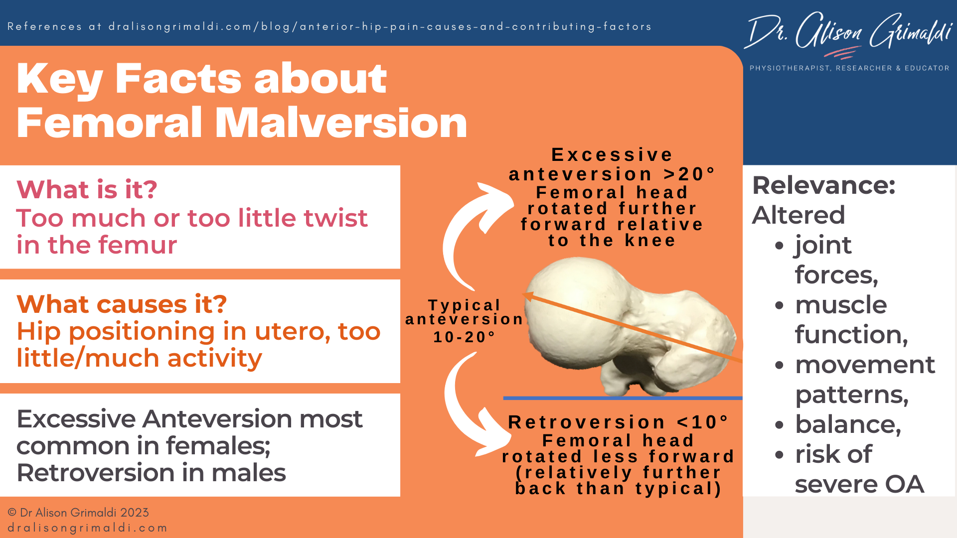
Femoral malversion refers to excessive femoral anteversion or femoral retroversion. Femoral version simply refers to the natural twist along the length of the femur between the femoral neck and the femoral condyles.
What is a typical amount of femoral version and what defines malversion?
There is no universally agreed degree of typical (normal) femoral version, but more recent studies have suggested some clearer guidelines.
- The typical human femur is anteverted somewhere between 10° and 20°
- Threshold for excessive femoral version: > 20° anteversion (malversion)
- Severe femoral anteversion: > 35° (severe malversion)
- Threshold for relative femoral retroversion: < 10° anteversion (malversion)
- Severe femoral retroversion: < 0° anteversion (severe malversion).
What causes femoral malversion?
Femoral anteversion is something that develops in utero, the typical femur of a newborn anteverted 30-40°, and then we spend our childhood gradually unwinding our femurs until the growth plates close in adolescence.
Development of femoral malversion appears to be related to:
- Development in utero – more common in breech babies.
- Sex – excessive anteversion more common in females; retroversion more common in males.
- Weightbearing stimulus – excessive anteversion more common in children with Cerebral Palsy; retroversion more common in obese adolescents.
- Physical activity – appears to stimulate derotation – low activity levels may result in excessive anteversion; high volume, 'hip-heavy' sports may accelerate derotation and result in retroversion.
- Muscle forces – abductors and flexors may decrease anteversion; adductors and extensors may increase anteversion.
- W-sitting?? Maybe in some children, associated with other factors.
Read more about the development and prevention of femoral malversion by clicking on the pink text.
What is the clinical relevance of femoral malversion?
Research around the impact of femoral malversion is still in its relative infancy, but there has been a growing interest in malversion in recent years.
Some key information we know so far about the impact of femoral malversion:
- Excessive femoral anteversion in children may result in intoeing gait, more frequent falls, earlier fatigue in weightbearing function and poorer balance than their peers.
- Excessive femoral anteversion increases hip joint contact forces in gait.
- Excessive femoral anteversion reduces abductor muscle capacity.
- Excessive femoral anteversion appears to be a risk factor for more severe hip OA.
- Femoral retroversion results in femoroacetabular impingement over a broader area, with potential for developing FAIS.
Read more about the impact of femoral malversion by clicking on the pink text.

Want to get better at assessing hip xrays, including assessment of femoral and acetabular shape?
Join Hip Academy and you will have immediate access to:
- A 6-page A4 PDF resource to download and use as a cheat-sheet when you are next looking at a patient’s images
- The recording of an exclusive 45-minute lecture & discussion on Deciphering Hip XRays, presented to the Hip Academy community.
- The Hip Academy Video Library of techniques, that includes 7 x 1-2 minute videos walking you through exactly how to complete key Xray measures within an image viewing system (PACS). Watch and copy the steps while assessing your patient’s images.
Muscle, movement and hip loading
There is currently inadequate prospective evidence that muscle or movement factors alone predispose to the development of anterior hip pain or pathology. It is likely that such impairments may have most impact when combined with other factors such as morphology, general health or iatrogenic factors. Muscle and movement impairments developed in response to injury or painful pathology may also contribute to worsening joint pain and pathology.13
We know that kinematics (movement patterns) and muscle activity both contribute to amount and direction of joint moments (kinetics). Changes in body or pelvic position, and gait parameters such as stride length can have a marked impact on joint loads.14,15
The action of deep hip muscles has been shown to reduce rim loading and change the direction of hip loading away from commonly damaged areas of acetabular cartilage (anterosuperior rim region).14 Good function may therefore be protective and dysfunction a potential contributor to altered joint forces.
The relationship between hip joint loading and joint health is an interesting topic.
As for any of our musculoskeletal tissues, there is probably a ‘sweet spot’ for hip joint loading somewhere in the middle, with both high loads and low loads potentially contributing to deterioration of hip joint health
The ‘use-it-or-lose-it’ principle remains an unwavering truth when it comes to musculoskeletal health and homeostasis. Volume, regularity and rate of increase of load imposed by physical activity may impact on tissue health.
Read more about the impact of alterations in physical activity and load management by clicking on the pink text.
With osteoarthritis, we most commonly talk about the relationships between high joint loads and cartilage deterioration, with links between the two established for knee OA.
However, at the hip there have been suggestions that underloading – reduced hip contact forces – is more of a problem than overloading. Diamond and colleagues argue that people with painful hip osteoarthritis walk with lower magnitude hip contact forces, in both end-stage and mild-moderate disease states.16
They suggest that it is this relative underloading that results in progressive hip joint degeneration, as the extra-cellular cartilage matrix requires adequate stimulus to maintain chondral health. Their solution, to increase loads by increasing stride length – a parameter shown to increase hip contact loads. 16
While it may seem tempting to apply this broadly, we must consider the finer detail about joint loads, including direction of forces – do we want to amplify problematic force vectors for that individual? Type of load is also a consideration for chondral health i.e., compressive load can be a great stimulus, while shear (sliding) force is not well tolerated by cartilage.
An individual’s morphology will strongly influence their intra-articular load profile. For example, aberrant small sliding movements of the femoral head in the acetabulum (microinstability) have been identified in those with cam type FAIS during gait, with implications for adverse impact on joint health over time.17 Those with acetabular dysplasia are also likely to experience greater shearing forces, contributing to their higher risk and earlier presentation of hip OA.
While we certainly need to embrace the concept of the possible role of joint underloading in progression and perhaps even as a contributor to development of degenerative change and for some, pain, the type and direction of load are also key considerations.
It will be interesting to see the results of further research in this space, exploring the relationships between these mechanical contributors to joint health and the pain experience.
I hope you’ve enjoyed this walk-through anterior hip pain causes and contributing factors. You’ll find plenty of other detail on this site, via the links throughout this blog.
Want to help more people who visit your clinic with anterior hip pain?
For assistance in applying this information in a clinical environment, I recommend you take my Anterior Hip and Groin Pain online course, or a practical workshop on this topic.

-
- A detailed examination of mechanisms of physical overload (morphology, movement, muscle) and impairments associated with anterior hip pain and groin pain, clinical diagnostic tests and management approaches.
- Including joint related-pain & bony impingement (intra & extra-articular), soft tissue-related pain and nerve-related pain.

- Perform diagnostic tests for anterior hip and groin pain and use that information for differential diagnosis of the most likely source of nociception or a primary clinical entity
- Perform tests that aim to elicit important information regarding potential contributors or drivers of the presenting condition.
- Determine the most appropriate management approach for an individual’s presenting condition.
Check Out Some More Relevant Blogs
References
- Heerey, J., Kemp, J., Mosler, A., et al. (2018). What is the prevalence of imaging-defined intra-articular hip pathologies in people with and without pain? A systematic review and meta-analysis. British Journal of Sports Medicine. 2018; 52(9):581-593.
- Martin RL, McDonough C, Enseki K, Kohreiser D, Kivlan BR. Clinical relevance of the ligamentum teres: a literature review. Int J Sports Phys Ther. 2019 Jun;14(3):459-467.
- Kaya M, Kano M, Sugi A. et al. Factors contributing to the failure of conservative treatment for acetabular labrum tears. Eur Orthop Traumatol. 2014;5:261–265.
- Maldonado DR, Laseter JR, Perets I, Ortiz-Declet V, Chen AW, Lall AC, Domb BG. The effect of complete tearing of the ligamentum teres in patients undergoing primary hip arthroscopy for Femoroacetabular Impingement and labral tears: A match-controlled study. Arthroscopy. 2019 Jan;35(1):80-88.
- Chaharbakhshi EO, Perets I, Ashberg L, Mu B, Lenkeit C, Domb BG. Do ligamentum teres tears portend inferior outcomes in patients with borderline dysplasia undergoing hip arthroscopic surgery? A match-controlled study with a minimum 2-year follow-up. Am J Sports Med. 2017 Sep;45(11):2507-2516.
- van Klij, P., Heerey, J., Waarsing, J. and Agricola, R., 2018. The Prevalence of Cam and Pincer Morphology and its Association with Development of Hip Osteoarthritis. Journal of Orthopaedic & Sports Physical Therapy, 48(4), pp.230-238.
- van Buuren M, Heerey J, Smith A et al. The association between statistical shape modeling-defined hip morphology and features of early hip osteoarthritis in young adult football players: Data from the femoroacetabular impingement and hip osteoarthritis cohort (FORCe) study. Osteoarthr Cartil Open. 2022;4(3):100275. doi:10.1016/j.ocarto.2022.100275
- Ahedi H, Winzenberg T, Bierma-Zeinstra S, Blizzard L, van Middelkoop M, Agricola R, Waarsing JH, Cicuttini F, Jones G. A prospective cohort study on cam morphology and its role in progression of osteoarthritis. Int J Rheum Dis. 2022 May;25(5):601-612.
- Wilkin GP, Ibrahim MM, Smit KM, Beaulé PE. A Contemporary Definition of Hip Dysplasia and Structural Instability: Toward a Comprehensive Classification for Acetabular Dysplasia. J Arthroplasty. 2017 Sep;32(9S):S20-S27.
- Henak CR, Ellis BJ, Harris MD, Anderson AE, Peters CL, Weiss JA. Role of the acetabular labrum in load support across the hip joint. J Biomech. 2011 Aug 11;44(12):2201-6.
- Gala L, Clohisy JC, Beaulé PE. Hip Dysplasia in the Young Adult. J Bone Joint Surg Am. 2016 Jan 6;98(1):63-73.
- Jacobsen JS, Hölmich P, Thorborg K, Bolvig L, Jakobsen SS, Søballe K, Mechlenburg I. Muscle-tendon-related pain in 100 patients with hip dysplasia: prevalence and associations with self-reported hip disability and muscle strength. J Hip Preserv Surg. 2017 Nov 17;5(1):39-46.
- Felson DT. Osteoarthritis as a disease of mechanics. Osteoarthritis Cartilage. 2013 Jan;21(1):10-5. doi: 10.1016/j.joca.2012.09.012. Epub 2012 Oct 4. PMID: 23041436; PMCID: PMC3538894.
- Meinders E, Pizzolato C, Gonçalves B, Lloyd D, Saxby D. and Diamond L. Activation of the deep hip muscles can change the direction of loading at the hip. Journal of Biomechanics. 2022; 135;111019.
- Lewis, C., Sahrmann, S. and Moran, D., 2010. Effect of hip angle on anterior hip joint force during gait. Gait & Posture, 32(4), pp.603-607.
- Diamond LE, Devaprakash D, Cornish B, Plinsinga ML, Hams A, Hall M, Hinman RS, Pizzolato C, Saxby DJ. Feasibility of personalised hip load modification using real-time biofeedback in hip osteoarthritis: A pilot study. Osteoarthr Cartil Open. 2021 Dec 25;4(1):100230. doi: 10.1016/j.ocarto.2021.100230. PMID: 36474469; PMCID: PMC9718151.
- Lewis CL, Uemura K, Atkins PR, Lenz AL, Fiorentino NM, Aoki SK, Anderson AE. Patients with cam-type femoroacetabular impingement demonstrate increased change in bone-to-bone distance during walking: A dual fluoroscopy study. J Orthop Res. 2023 Jan;41(1):161-169. doi: 10.1002/jor.25332. Epub 2022 Apr 6. PMID: 35325481; PMCID: PMC9508282.

